Fig. 1
Fig. 1
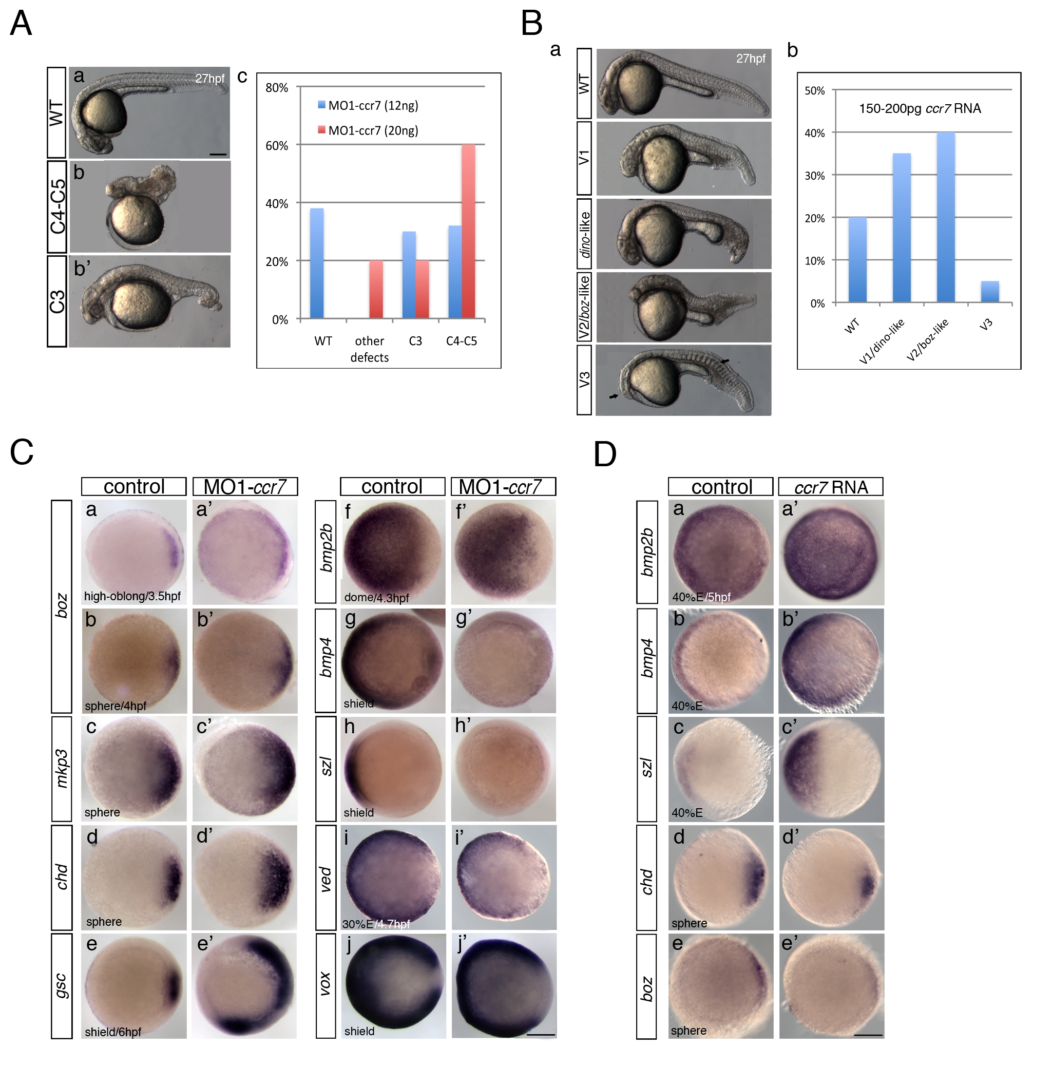
Ccr7 is required for proper AP and DV embryo patterning.
(A) The spectrum of dorsalized phenotypes in embryos injected with MO1-ccr7 at 27 hpf, ranging from highly dorsalized (C4–5; b) with truncated tails and trunks to C3 with tail deficiencies (b2) (total n = 190, seven independent experiments for 12 ng MO1-ccr7 and n = 35, one experiment for 20 ng MO1-ccr7). The frequency of each phenotypic category is indicated in the right panel (c). The scale bars represent 200 μm in all figures. (B) Ccr7 overexpression (150–200 pg RNA) caused a spectrum of ventralized phenotypes, ranging from V3 to V1 (arrows show anterior and notochord deficiencies) to WT-like (a). The frequency of each phenotypic category is indicated in the right panel (b; n = 96, two experiments). (C) Expression of dorsal/ventral markers in Ccr7 morphants compared to control embryos revealed by WISH. (a–e2) Expression of dorsal genes was upregulated or expanded: at high-oblong stage (3.3–3.7 hpf), boz, n = 8/8; at sphere stage (4 hpf), boz, n = 16/28; mkp3, n = 9/12; chd, n = 11/15; at shield stage (6 hpf), gsc, n = 18/37. (f–j2) Expression of ventral genes was reduced: at dome (4.3 hpf) stage, bmp2b, n = 19/24; at 30% epiboly stage (4.7 hpf), ved, n = 12/14; at shield stage, bmp4, n = 9/10; szl, n = 11/12; vox, n = 16/22. Animal views, dorsal to the right. (D) Expression of dorsal/ventral markers in Ccr7-overexpressing embryos revealed by WISH. Expression of ventral markers was expanded (a–c2), while dorsal markers were decreased (d–e2): at sphere stage, boz, n = 8/8; chd, n = 10/13; at 40% epiboly stage (5 hpf), bmp2b, n = 12/12; bmp4, n = 14/14; szl, n = 13/13. Animal views, dorsal to the right.

