
|
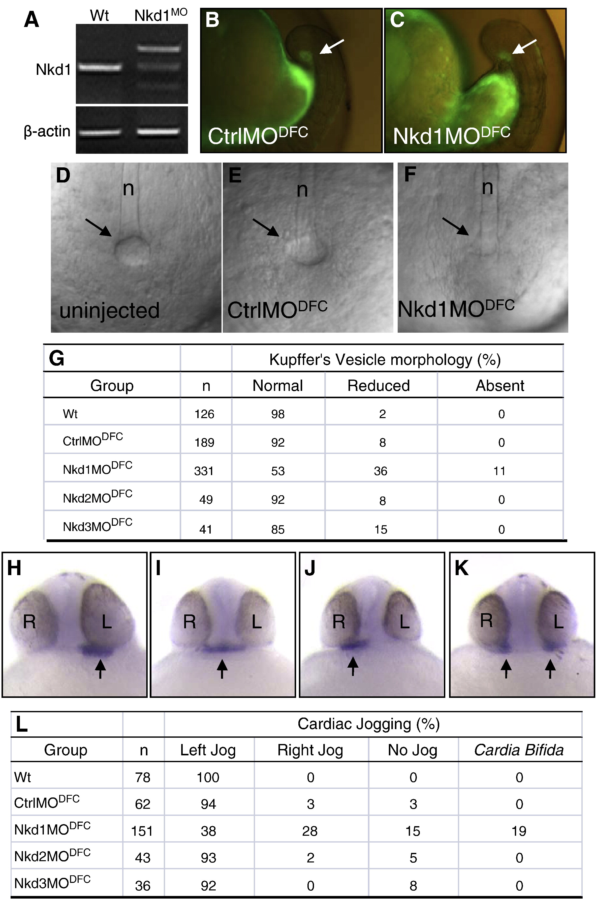
Fig. 2 Heart laterality defects after Nkd1 knockdown in DFCs. (A) RT-PCR using cDNA from wt or Nkd1MO-injected embryos: β-actin or Nkd1 were amplified. (B and C) Arrows denote efficient targeting of CtrlMO and Nkd1MO into the DFCs. (D–F) Bright field image of KV in wt (D), CtrlMODFC (E) and Nkd1MODFC(F) embryos, at 10-somite stage. Arrows indicate vesicle location; n = notochord. (G) Summary of KV morphology defects. (H–K) Arrows denote expression of nkx2.5 in the heart, on the left side in wt (H) and on the middle (I), right (J) and bilaterally (K) in Nkd1MODFC embryos. (L) Summary of cardiac jogging defects.
Reprinted from Developmental Biology, 348(1), Schneider, I., Schneider, P.N., Derry, S.W., Lin, S., Barton, L.J., Westfall, T., and Slusarski, D.C., Zebrafish Nkd1 promotes Dvl degradation and is required for left-right patterning, 22-33, Copyright (2010) with permission from Elsevier. Full text @ Dev. Biol.