IMAGE
Fig. S5
Image
Figure Caption
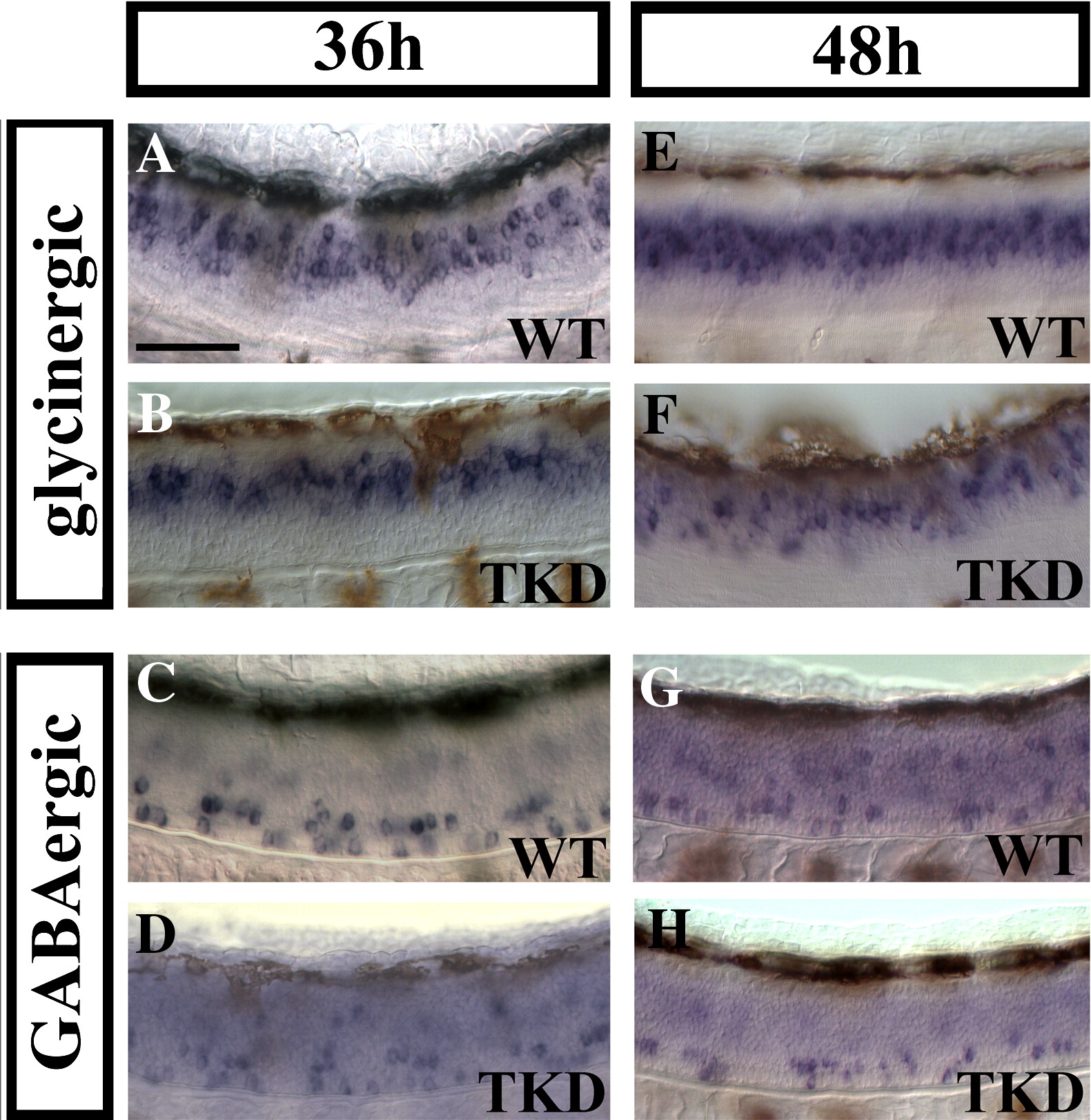
Fig. S5 Triple knock-down embryos still have reduced numbers of glycinergic and GABAergic spinal neurons at 36 h and 48 h. Lateral views of in situ hybridisation for markers of glycinergic (A, B, E and F) and GABAergic (C, D, G and H) cell fates in wild-type (WT; A, C, E and G) and triple knock-down (TKD; B, D, F and H) trunks at 36 h (A–D) and 48 h (E–H). (Cell counts for this data are shown in Supp. Data Figs. 4A and A′.) Brown cells are melanophores that have differentiated and started to migrate by these stages. Rostal is left, dorsal is top. Scale bar = 50 μm.
Figure Data
Acknowledgments
This image is the copyrighted work of the attributed author or publisher, and
ZFIN has permission only to display this image to its users.
Additional permissions should be obtained from the applicable author or publisher of the image.
Reprinted from Developmental Biology, 323(1), Batista, M.F., and Lewis, K.E., Pax2/8 act redundantly to specify glycinergic and GABAergic fates of multiple spinal interneurons, 88-97, Copyright (2008) with permission from Elsevier. Full text @ Dev. Biol.

