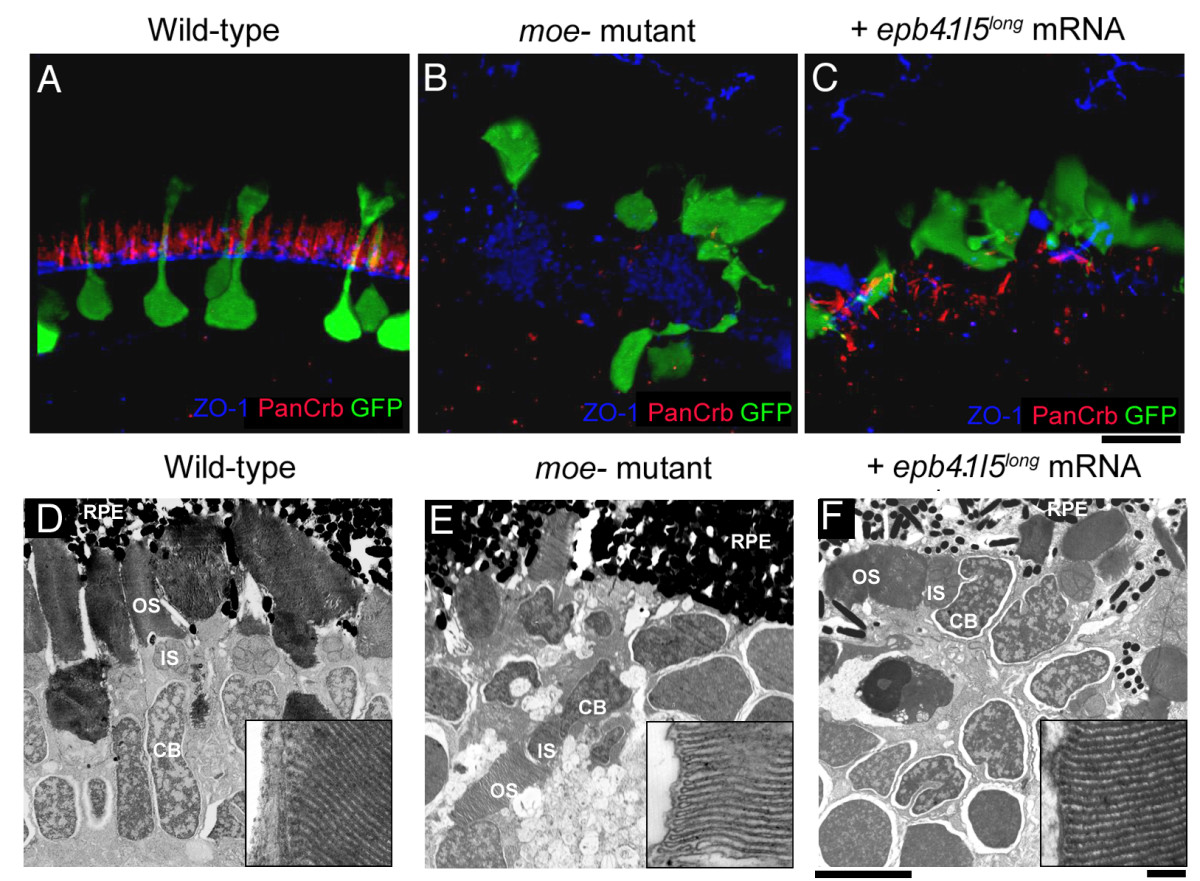
Fig. 6 The outer limiting membrane (OLM) is not restored in moe- mutants by injection of epb4.1l5long mRNA. (A) In wild-type larvae at 6 dpf, Crumbs proteins localize just apical to the OLM, which is labeled by anti-ZO-1. (B) In moe- mutants, very little Crumbs protein is visible and ZO-1 labeling is disorganized. (C) In epb4.1l5long mRNA injected moe- mutants, streaks of panCrb labeling are visible, but an organized OLM is absent. Ultrastructural transmission electron microscopic analysis at 6 dpf in wild-type (D), moe- mutants (E) and epb4.1l5long mRNA injected moe- mutants (F) retinas. Electron dense outer segments are seen in all individuals. Insets, higher magnifications of rod outer segments showing regular disc stacking is present in all individuals (100,000X). RPE, retinal pigmented epithelium; OS, outer segments; IS, inner Segments; CB, cell body. (A-C) are confocal z-projections, scale bar 10 μm (D-F). Scale bars, 5 μm (D-F), 100 nm (insets).
Image
Figure Caption
Figure Data
Acknowledgments
This image is the copyrighted work of the attributed author or publisher, and
ZFIN has permission only to display this image to its users.
Additional permissions should be obtained from the applicable author or publisher of the image.
Full text @ BMC Dev. Biol.

