Fig. 6
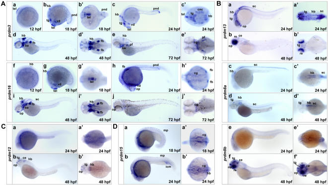
Fig. 6 Nervous system-expressed SET domain genes. (A) Expression patterns of closely related prdm3 and prdm16. (a–j) Lateral views (anterior to the left) of embryos at 12, 18, 24, 48 and 72 hpf. (b′ and g′) Ventral views of the embryos in b and g. (c′–e′ and h′–j′) Dorsal views of the embryos in c–e and h–j. Note the partially overlapping expression of prdm3 and prdm16. (B) Expression patterns of prdm13, prdm8a and prdm8b. (a–f) Lateral views (anterior to the left) of embryos at 24 and 48 hpf. (a′–f′) Dorsal views of the embryos in a–f. Note that the expression of prdm8a is mostly restricted in hindbrain and spinal chord (c, d and c′, d′), whereas that of prdm8b is restricted in olfactory placode, tegmentum, cerebellum and retina (f and f′). (C) Expression pattern of prdm12. (a and b) Lateral views (anterior to the left) of embryos at 18, 24 hpf. (a′ and b′) Dorsal views of the embryos in a and b. At 48 hpf, prdm12 is expressed in olfactory placode, tegmentum, cerebellum and hindbrain. (D) Expression pattern of prdm15. (a and b) Lateral views (anterior to the left) of embryos at 18, 22 hpf. (a′ and b′) Dorsal views of the embryos in a and b. Note that prdm15 is expressed in cranial ganglia neurons (a′ and b′) as well as in muscle pioneer cells and intermediate cell mass (a and b). ba, branchial arches; ce, cerebellum; cg, cranial ganglia; cnc, cranial neural crest; fb, fin buds; hb, hindbrain; icm, intermediate cell mass; mp, muscle pioneer; op, olfactory placode; pnd, pronephric duct; re, retina; sc, spinal chord; tel, telencephalon; tg, tegmentum; vd, ventral diencephalons.

