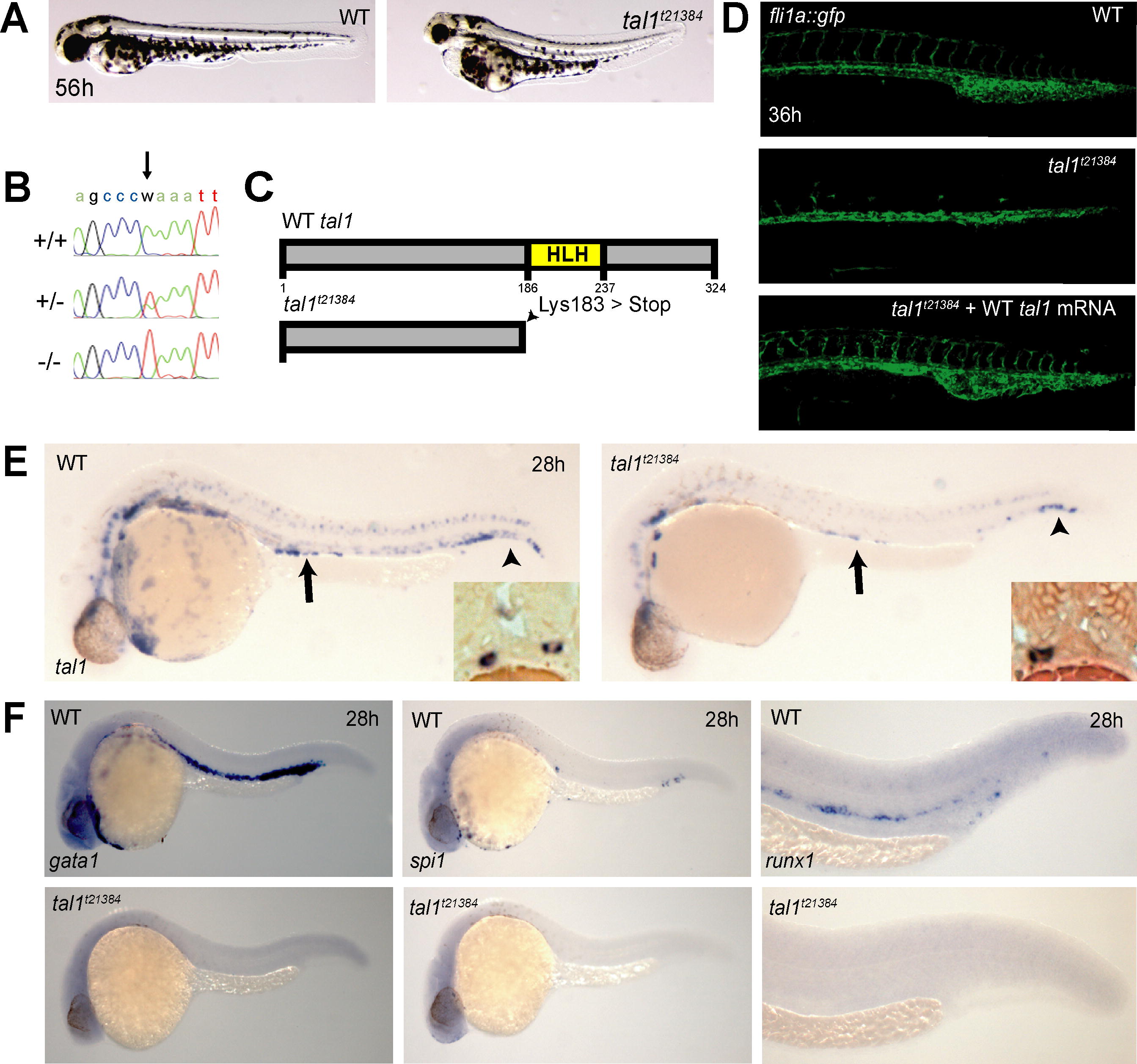
Fig. 1 A Truncating Mutation in the Zebrafish tal1 Gene (A) Live micrographs of wt and tal1t21384 mutant embryos at 56h. Mutant embryos have slightly curved tails, smaller eyes, and pericardial edema. (B) Electropherograms of wt (+/+), heterozygous (+/-) and tal1t21384 homozygous genomic DNA from bp 7–17 of the third tal1 coding exon. The A→T transversion (arrow) mutates lysine 183 to an ochre (TAA) stop codon (K183X). (C) Schematic diagram of the primary Tal1 protein structure. The DNA-binding bHLH domain is indicated in yellow. The tal1t21384 mutation deletes the Tal1 protein C terminus including the complete helix-loop-helix domain (D) The tal1t21384 mutation leads to severe defects in intersomitic vessel formation at 36 hpf, visualized in a fli1a:gfp transgenic background. Early endothelial pattering can be rescued by injecting 20 pg wt tal1 mRNA into mutant embryos. (E) Loss of tal1 expression in tal1t21384 mutant embryos. In situ hybridization for tal1 shows a loss of tal1 expression in erythroid cells and a reduction of tal1 expression in the spinal chord. tal1 expression is retained at normal levels in some hematopoietic/endothelial progenitor cells in the tail (arrowheads) and in some mesenchymal cells of unknown nature just dorsal to the yolk extension (arrows). These bilateral cell populations lie ventral to the pronephric tubules and lateral to the gut endoderm (insets). Also note the loss of tal1 expression in the ventral wall of the dorsal aorta containing, in wt embryos, the definitive hematopoietic stem cells. (F) Loss of hematopoiesis in tal1t21384 mutant embryos. In situ hybridization for hematopoietic markers at 26 hpf shows a loss of primitive erythroid cells (gata1), primitive myeloid cells (pu.1), and definitive hematopoietic stem cells (runx1).
Image
Figure Caption
Figure Data
Acknowledgments
This image is the copyrighted work of the attributed author or publisher, and
ZFIN has permission only to display this image to its users.
Additional permissions should be obtained from the applicable author or publisher of the image.
Full text @ PLoS Genet.

