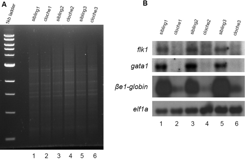
Fig. 2 Analysis of amplified cDNA quality. A: DNA electrophoresis analysis. A total of 100 ng of each amplified cDNAs derived from the single 18-somite clo homozygous mutant (lanes 2, 4, and 6) and wild-type or heterozygous sibling embryos (lanes 1, 3 and 5) was separated on 1% agarose gel. Ethidium bromide staining shows an evenly distributed smear of DNA in each lane. B: Virtual Northern analysis. cDNAs of A were transferred to a Hybone-N+ membrane and probed with digoxigenin-labeled flk1, gata1, e1-globin and actin. Whereas elf1a transcript was maintained at a similar level in all the samples, expression of flk1, gata1, and e1-globin were either absent or drastically reduced in the clo mutant embryos (lanes 2, 4, and 6).
Image
Figure Caption
Figure Data
Acknowledgments
This image is the copyrighted work of the attributed author or publisher, and
ZFIN has permission only to display this image to its users.
Additional permissions should be obtained from the applicable author or publisher of the image.
Full text @ Dev. Dyn.

