
Fig. 8
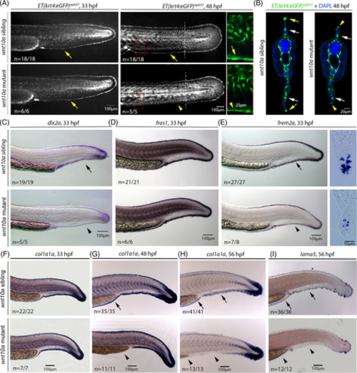
MFF collapse in wnt10a mutant MFF involves the down-regulation of dlx2a and crucial ECM protein-encoding genes in distal MFF cells. (A) At 33 hpf (left panels), before MFF collapse becomes morphologically visible, wnt10a mutant displays unaltered expression of the Et(krt4:eGFP)sqet37 transgene both in fin mesenchymal cells and in distal-most epidermal cells of the MFF (indicated by yellow arrows). At 48 hpf (middle panels and right panels for enlarged views of regions framed in middle panels), when MFF collapse is obvious and in progress, fin mesenchymal cells of the mutant still show normal Et(krt4:eGFP)sqet37 expression, whereas transgene expression in distal epidermal cells is reduced (indicated by yellow arrowheads) in comparison to wild-type sibling (indicated by yellow arrow). (B) Transverse sections of 48 hpf Et(krt4:eGFP)sqet37 transgenics at the location indicated in middle panels of (A) by dashed lines, indicating the normal positioning and transgene expression in fin mesenchymal cells (white arrows), but the reduced transgene expression in distal epidermal cells both in the collapsing dorsal and ventral MFF of the mutant (yellow arrowheads) compared to its sibling (yellow arrows). (C–E) In situ hybridization indicates that at 33 hpf, before MFF collapse becomes morphologically visible, the expression of the distal marker gene dlx2a is strongly down-regulated in distal-most epidermal cells of the ventral MFF (C). Expression of fras1, a marker for distal-most cleft cells,65 appears unaltered in mutants compared to their wild-type siblings (D), whereas expression of frem2a, a marker for distal ridge cells,65 is reduced, as also obvious in transverse Durcupan sections (E). (F–I) In situ hybridization indicates that at 33 hpf (left panels), before MFF collapse becomes morphologically visible, expression of col1a1a, encoding an essential component of actinotrichia, is normally expressed in distal-most epidermal cells of the MFF of wnt10a mutants (F), while expression levels become progressively reduced at 48 hpf ((G); most obvious in minor lobe) and at 56 hpf (H), when fin collapse is in progress. A similar progressive reduction of expression in the MFF of wnt10a mutant is observed for lama5, encoding an essential component of the MFF basement membrane ((I) for 56 hpf; earlier stages not shown). Arrows indicate regular expression; arrowheads indicate less or non-detectable expression. ECM, extracellular matrix; MFF, median fin fold. |