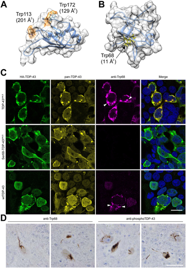
Trp68 is exposed in cytoplasmic TDP-43 aggregates in cultured cells and in sporadic ALS motor neurons.A, representative centroid structure of the RRM1 domain of TDP-43, obtained from equilibrium molecular dynamics (MD) simulation. Trps 113 and 172 are significantly exposed compared to most native structures in the protein databank (11). B, representative centroid structure of the N-terminal domain of TDP-43, obtained from equilibrium molecular dynamics simulation. Trp68 is nearly fully buried: only about 9% of its tripeptide Gly-Trp-Gly value of 247 Angstrom2 is exposed in the centroid structure. C, the affinity-purified rabbit polyclonal anti-Trp68 antibody (red) was tested for reactivity and specificity in cells transfected with different TDP-43 constructs, then fixed and stained 48 h later. A mouse pan-TDP-43 antibody against the C-terminal domain and a chicken anti-HA-tag antibody were used to test co-localization with TDP-43 (yellow) and HA-TDP-43 (green), respectively. The anti-Trp68 antibody specifically recognizes mislocalized cytoplasmic TDP-43 aggregates in TDP-43ΔNLS-transfected cells but not those lacking Trp68 (Ser68 TDP-43ΔNLS) nor does it recognize nuclear TDP-43. The composite images are a merge between nuclear, anti-Trp68, and anti-HA-tag staining. Scale bar represents 20 μm. D, representative images of TDP-43 pathology in ALS cervical spinal cord sections from two subjects immunostained with anti-Trp68 in paraffin-embedded tissue. The left panel shows the anti-Trp68 antibody and right the commercial pTDP-43 antibody. Motor neurons show thread-like inclusions using the anti-Trp68 antibody and the commercial pTDP-43 antibody, but no immunoreactivity is observed of natively folded TDP-43 in neuronal or glial nuclei Scale bar represents 50 μm. ALS, amyotrophic lateral sclerosis; RRM, RNA recognition motif; TDP, TAR DNA-binding protein.
|
