FIGURE

Figure 3.

ID
ZDB-FIG-240509-65
Publication
Guo et al., 2024 - Zebrafish Mbd5 binds to RNA m5C and regulates histone deubiquitylation and gene expression in development metabolism and behavior
Other Figures
All Figure Page
Back to All Figure Page
Figure 3.

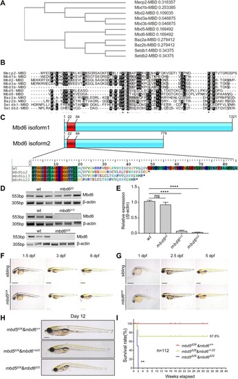
CRISPR/Cas9-mediated germ-line disruption of Mbd6, a MBD protein closely related to Mbd5, reveals redundant function in larval growth and physiology. (A) Phylogenetic analysis of the MBD domain-containing proteins. (B) Protein alignment of the MBD domain. Asterisks represent identical amino acids among the members. Periods indicate that the amino acid is identical or similar across the members. (C) Schematic analysis of the protein variants of Mbd6 and predicted amino acid sequence of wild type and different alleles of mbd6 mutants. (D) Semi-quantitative RT-PCR analysis reveals the mbd6 mRNA expression in mbd6Δ2, mbd6Δ13 and mbd6Δ22 alleles. β-actin was used as the internal control. (E) Statistical analysis of the mbd6 mRNA expression in mbd6Δ2, mbd6Δ13 and mbd6Δ22 alleles. Value in each column represents mean ± SD, ns, no significance, ****P< 0.0001. (FG) Whole-mount lateral view of mbd5Δ29 and mbd6Δ22 mutants at indicated developmental stages under bright field condition. At least 10 were examined for each condition. (H) Lateral view of mbd5Δ29& mbd6Δ22 double mutants and those siblings at 12 dpf. N = 48 (mbd5Δ29& mbd6+/+, N = 12; mbd5Δ29& mbd6+/Δ22, N = 28; mbd5Δ29& mbd6Δ22, N = 8). (I) Survival analysis of the progeny of mbd5ΔMBD& mbd6+/Δ22 in cross during 32 weeks after birth. N = 112, Log-rank (Mantel–Cox) test, ** P< 0.01. Scale bar: 0.5 mm (F, G).

Expression Data

Expression Detail
Antibody Labeling
Phenotype Data

Phenotype Detail
Acknowledgments
This image is the copyrighted work of the attributed author or publisher, and ZFIN has permission only to display this image to its users. Additional permissions should be obtained from the applicable author or publisher of the image. Full text @ Nucleic Acids Res.