|
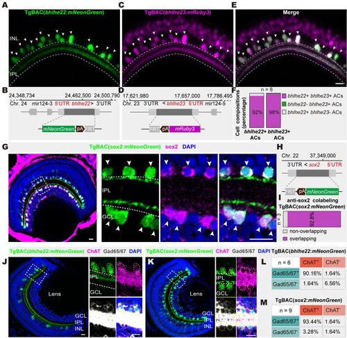
Genetically marking 2 GABAergic/cholinergic AC types. (A and B) Representative image (A) of derived transgenic line and schematic (B) of BAC construct design of bhlhe22. (C and D) Representative image (C) of derived transgenic line and schematic (D) of BAC construct design of bhlhe23. (E) Merged image of (A) and (C) after crossing transgenic lines TgBAC(bhlhe22:mNeonGreen) with TgBAC(bhlhe23:mRuby3). (F) Cell composition analysis of bhlhe22 and bhlhe23 labeling cells in (E). Larvae used for composition analysis are from TgBAC(bhlhe22:mNeonGreen,bhlhe23:mRuby3), n = 8. (G) Immunostaining of sox2+ ACs in the transgenic fishline TgBAC(sox2:mNeonGreen). Solid white arrow head indicated colocalization of sox2 transgenic fishline and SOX2 antibody. (H) Schematic of BAC construct design of TgBAC(sox2: mNeonGreen). (I) The bar plot showing that the majority of sox2+ ACs in the transgenic fishline is colocalized with SOX2 antibody. (J and K) Images showing the colabeling of bhlhe22 + type (J, green) and sox2+ type (K, green) with ChAT (magenta) and Gad65/67 (gray). (L and M) Quantification in (J and K). Images above are captured from 5-dpf larval fish. Dashed yellow circles, ACs with positive signals. The data underlying this figure can be found in S3 Data. Scale bars, 10 μm. AC, amacrine cell; BAC, bacterial artificial chromosome; ChAT, choline acetyltransferase; dpf, days post-fertilization; GCL, ganglion cell layer; INL, inner nuclear layer; IPL, inner plexiform layer.
|