
Fig. 8
- ID
- ZDB-FIG-231215-8
- Publication
- Tu et al., 2022 - Dhx38 regulates the maintenance and differentiation of erythro-myeloid progenitors and hematopoietic stem cells by alternative splicing
- Other Figures
- All Figure Page
- Back to All Figure Page
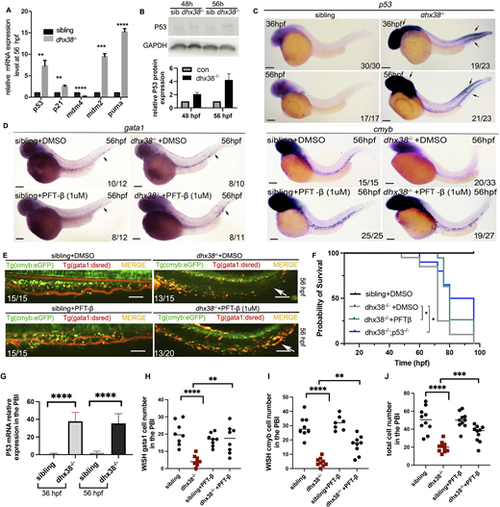
EMPs and HSPCs in the dhx38 mutants undergo P53-dependent apoptosis. (A) qRT-PCR analysis of the genes involved in P53 signaling shows a significant increase in the expression of p53, p21, puma, mdm4 and mdm2. n≥12 per group. Experiments were performed with three replicates; gapdh was used as an internal control. (B) Western blotting analysis shows an increased expression of P53 in dhx38−/− zebrafish at 48 hpf, which increases further at 56 hpf. (C) The expression pattern of p53 detected by WISH reveals that P53 begins to accumulate in the PBI at 36 hpf and 56 hpf. Black arrows indicate the expression of p53. (D) Decreased expression of cmyb (right) and gata1 (left) in the dhx38 mutants can be rescued following incubation with the P53 inhibitor PFT-β (1 µM). Black arrows indicate the expression of gata1. (E) In vivo imaging of hematopoietic progenitor cells in Tg(cmyb:eGFP;gata1:dsred) at 56 hpf. The overall number of hematopoietic progenitor cells in the PFT-β group was higher than in the DMSO group. White arrows indicate blood cells in the CHT region. (F) The survival curve shows that PFT-β and dhx38−/−;p53−/− rescue the survival rate of dhx38 mutants. Compared with dhx38−/− embryos, the survival ratios of dhx38−/− embryos incubated with PFT-β at 72 hpf (**P=0.003) and 76 hpf (*P=0.079) are significant. Compared with dhx38−/− embryos, the survival ratios of dhx38−/−;p53−/− at 72 hpf (*P=0.012), 76 hpf (*P=0.029) and 80 hpf (**P=0.007) are significant. n≥20 per group. Experiments were performed with three replicates; (G-J) Quantification of cells from C-E. Data show the mean±s.d. Significance was determined with two-tailed unpaired Student's t-test. *P<0.05; **P<0.01; ***P<0.001; ****P<0.0001. All scale bars: 200 μm. |