|
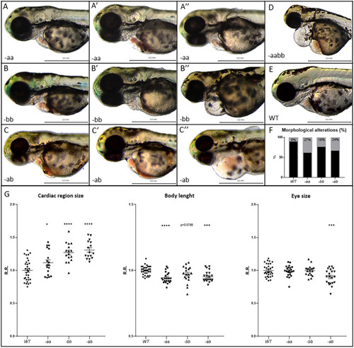
Cardiac alterations and developmental delay in Dsp mutant lines. A–F Mutant embryos (A–C″, D) display cardiac alterations, compared to WT (E). Homozygous -aa (A–A″) and -bb (B–B″) mutant hearts appear dilated and/or structurally altered, with cardiac pericardial effusion and/or hemopericardium in the cardiac region. Double heterozygotes -ab (C–C″) show a more serious phenotype than -aa and -bb lines. Double homozygotes -aabb display the most severe phenotype (D). F Percentage of heart alteration in different genotypes. Sample size: n = 100. G Cardiac size analysis shows that all mutants present heart dilation. Body length and eye size measurements indicate developmental delay in mutants, especially in the -ab condition. All embryos are at 3 dpf in lateral view, anterior to the left. Sample size Cardiac region size: WT n = 31; -aa n = 18; -bb n = 16; -ab n = 16. Sample size Body length: WT n = 32; -aa n = 22; -bb n = 19; -ab n = 21. Sample size Eye size: WT n = 32; -aa n = 22; -bb n = 19; -ab n = 21. R.R. relative ratio. Error bars: SEM. *p < 0.05; **p < 0.01; ***p < 0.001; ****p < 0.0001. Test: One-way ANOVA followed by Tukey’s test.
|