
Fig. 6
- ID
- ZDB-FIG-231208-26
- Publication
- Higashi et al., 2023 - Zinc-based Ultrasensitive Microscopic Barrier Assay (ZnUMBA): a live imaging method to detect local barrier breaches
- Other Figures
- All Figure Page
- Back to All Figure Page
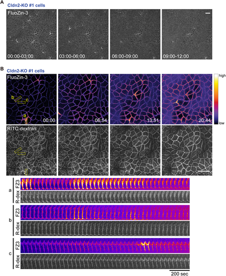
ZnUMBA detects naturally occurring leaks at cell–cell boundaries. (A) Brightest-point projections of FZ3 signals using Cldn2-KO cells over four time intervals (min:s). Scale bar: 20 µm. (B) Time-lapse images (min:s) of ZnUMBA using Cldn2-KO cells. RITC–dextran (lower panels, white) was added to the FZ3 solution to visualize the basal compartment of paracellular space. The Fire lookup table from ImageJ has been applied to the FZ3 channel (upper panels). Kymographs of the FZ3 and RITC–dextran (R-dex) signals in the yellow rectangles (a, b and c) are shown (bottom). Note that ZnUMBA signal fluctuates over time, whereas RITC–dextran signal remains unchanged. Scale bar: 20 µm. See also Movie 2. Images in A and B are representative of three experiments. |