
Fig. 2
- ID
- ZDB-FIG-231208-22
- Publication
- Higashi et al., 2023 - Zinc-based Ultrasensitive Microscopic Barrier Assay (ZnUMBA): a live imaging method to detect local barrier breaches
- Other Figures
- All Figure Page
- Back to All Figure Page
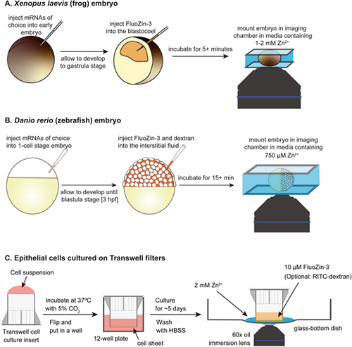
Modification of ZnUMBA for different model systems. (A) X. laevis embryos are injected with mRNAs of interest at early embryo stages (1–4-cell stage). After developing to gastrula stage, FZ3 is injected into the blastocoel, and the embryos are incubated for a minimum of 5 min to allow the injection site injury to heal. Finally, just prior to imaging, embryos are mounted in Zn2+-containing medium and imaged via confocal microscopy. (B) Zebrafish embryos are injected with mRNAs of interest at the 1-cell stage. Embryos are dechorionated, and after developing to 3 hpf (1k-cell stage), embryos are injected with FZ3 and fluorescently labeled dextran into the interstitial fluid. Embryos are incubated for ∼15 min to allow the injection site injury to heal. Finally, embryos are mounted in low-melting-point agarose, and Zn2+-containing medium is added before the start of imaging. (C) Experimental setup for ZnUMBA using MDCK II cultured epithelial cells. The Transwell filter cup is placed upside-down on a clean surface, and 1×10⁵ cells resuspended in 300 µl of DMEM are seeded onto the bottom surface of the filter. The filter is incubated at 37°C in a moist CO2 incubator for 10–14 h. After cells are attached to the surface, the filter cup is inverted and placed into a well of a 12-well plate. The cells are cultured for ∼5 d until the TER increases. For ZnUMBA, 500 µl of HBSS containing 2 mM ZnCl2 is placed on the glass-bottom dish, and the Transwell filter cup with the cell sheet attached is placed onto the Zn2+-containing medium. HBSS containing 10 µM FZ3 and 1 µM CaCl2-EDTA is added into the filter cup (upper compartment), and the fluorescence is observed using an inverted fluorescence microscope. For visualization of the basal compartment, RITC–dextran can be included in the ZnCl2 solution. |