Fig. 1
- ID
- ZDB-FIG-231201-59
- Publication
- Fu et al., 2023 - HSP90β prevents aging-related cataract formation through regulation of the charged multivesicular body protein (CHMP4B) and p53
- Other Figures
- All Figure Page
- Back to All Figure Page
|
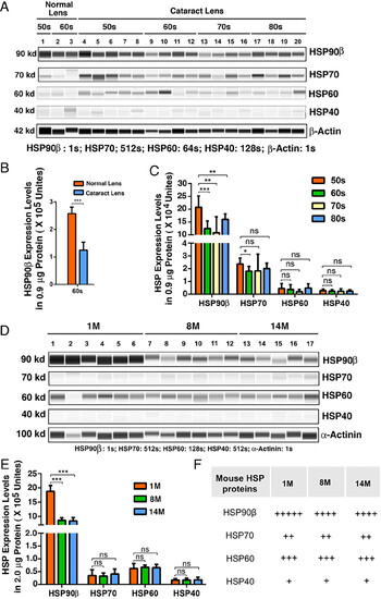
HSP90β is the most dominant HSP in normal human and mouse lenses and becomes significantly down-regulated during human cataractogenesis and aging of mouse lens. (A) The automated western immunoblot (WES) analysis of HSP90β, HSP70 (the HSP70 antibody recognizing both inducible and constitutive isoforms), HSP60 and HSP40 between normal lens and cataract patients in different age groups. Output western blot style data of four HSPs and the β-Actin (as control) with the exposure time indicated. Each lane was loaded 0.9-μg protein. (B) Quantification data derived from the software-calculated average of seven exposures (1-512 s). The quantification results show that HSP90β is significantly down-regulated from normal human lens to cataractous lens of the same age group (60s group including 61- to 69-y-old subjects). (C) Quantification results show the differential expression levels of four HSPs among different age groups of cataract patients (see SI Appendix, Tables S2–S5 for details of different age groups). (D) The WES analysis of HSP90β, HSP70 (the HSP70 antibody recognizing both inducible and constitutive isoforms), HSP60 and HSP40 in mouse lenses of 1-, 8-, and 14-mo-old animals. Output western blot style data of four HSPs and the α-Actinin (as control) with the exposure time indicated. Each lane was loaded 2-μg protein. (E) Quantification data derived from the software-calculated average of seven exposures (1-512s). The quantification results show that HSP90β is significantly down-regulated from normal mouse lens to aged mouse lens (8- to 14-mo). In contrast, other three HSPs display no age-dependent changes. (F) Summary of the WES analysis results of four HSPs in normal and aging mouse lenses. +++++, ++++, +++, ++ and + refer to expression levels of 1 × 10⁶, 1 × 10⁵ to 1 × 10⁶, 5 × 10⁴ to 1 × 10⁵, 3 × 10⁴ to 5 × 10⁴, and less than 3 × 10⁴ units, respectively, in 2-µg total protein. Error bar represents SD. *P < 0.05; **P < 0.01; ***P < 0.001; ns, statistically not significant. |

