|
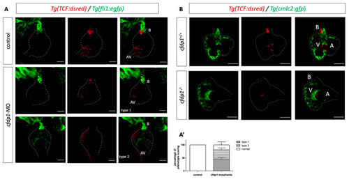
cfdp1 abrogation shows impaired Wnt/β-catenin signaling. (A) Confocal images of cfdp1 morphants show that Wnt/β-catenin reporter activity is diminished compared to the uninjected sibling controls. Max projection of z-stack confocal images of 120 hpf cfdp1-MO embryos. Endothelial cells are labeled with green (Tg(fli1:EGFP)) and Wnt-activated cells are labeled with red (Tg(7xTCF-Xla.Siam:nlsmCherry)); n = 6 in each of three independent experiments. (A’) percentage of phenotypic scoring. AV, atrioventricular valve. B, bulbus arteriosus. Scale bar 150 μm. (B) Confocal images of 120 hpf cfdp1 mutant and wild-type siblings expressing nlsmCherry in Wnt-activated cells and Tg(cmlc2:eGFP) in all cardiomyocytes (Ν = 3, n = 13). Scale bar: 50 μm.
|