FIGURE

Fig. 1

ID
ZDB-FIG-230707-47
Publication
Zhang et al., 2023 - Wdr5-mediated H3K4me3 coordinately regulates cell differentiation, proliferation termination, and survival in digestive organogenesis
Other Figures
All Figure Page
Back to All Figure Page
Fig. 1

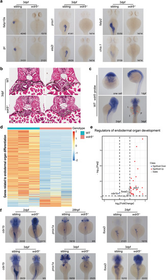
The differentiation of intestine, liver, and exocrine-pancreas is impaired in wdr5/− mutant embryos

a Whole-mount in situ hybridization (WISH) of wild type (WT) and wdr5/− mutant embryos at 3 dpf to show the expression of differentiation genes for different digestive organs including: the fabp10a and gc for liver, prss1and ela2l for exocrine pancreas, fabp2 and chia.1 for intestine as indicated. b Hematoxylin-Eosin (H&E) staining of cryosections to show liver (L), intestine (I) and exocrine pancreas (P) in WT and wdr5−/− mutant embryos at 3 dpf. Scale bar: 40 μm. c WISH of wdr5 in WT at one cell stage, 1, 2 and 3 dpf. d Heatmap of genes related to endodermal organ differentiation in WT and wdr5−/− mutant embryos at 3 dpf. e Volcano plot to show differential expressed regulators of endodermal organ development between WT and wdr5/− mutant embryos at 3 dpf (wdr5−/− mutant vs WT, |log2FoldChange | ≥ 1, Padj < 0.05). f WISH of WT and wdr5−/− mutant embryos at different time points with different digestive organ development regulators including: pan-endodermal marker foxa3, hepatic marker prox1a and intestine marker cdx1b as indicated. n indicates the number of zebrafish embryos in each group.

Expression Data

Expression Detail
Antibody Labeling
Phenotype Data

Phenotype Detail
Acknowledgments
This image is the copyrighted work of the attributed author or publisher, and ZFIN has permission only to display this image to its users. Additional permissions should be obtained from the applicable author or publisher of the image. Full text @ Cell Death Discov