Fig. 1
- ID
- ZDB-FIG-230404-17
- Publication
- Leslie et al., 2022 - Biallelic DAW1 variants cause a motile ciliopathy characterized by laterality defects and subtle ciliary beating abnormalities
- Other Figures
- All Figure Page
- Back to All Figure Page
|
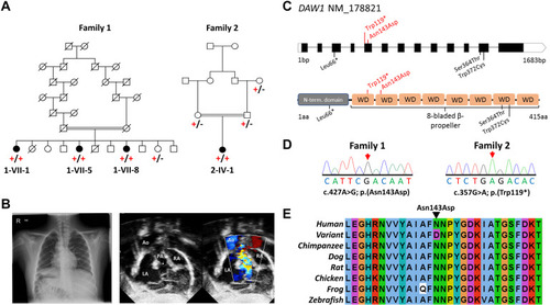
Figure 1. DAW1 variants, family pedigrees, and clinical images. A. Simplified pedigree of the extended Mennonite family (family 1) and the Palestinian family (family 2) in which segregation of the [NM_178821.2:c.427A>G; p.(Asn143Asp)] and [NM_178821.2: c.357G>A; p.(Trp119∗)] DAW1 variants are indicated (+, variant; –, wild type). B. Chest radiograph from individual 2-IV-1 from family 2 showing the right sided aortic arch, right sided stomach, and situs inversus. Echocardiogram imaging from the same individual showing atrial situs inversus. C. Schematic showing DAW1 intron–exon genomic organization (upper panel) and polypeptide domain architecture (lower panel), with position of DAW1 variants indicated. Variants identified in this study are indicated above the schematic in red, variants identified in previous studies are indicated below the schematic in black. D. Sequencing electropherograms from individuals homozygous for the p.(Asn143Asp) and p.(Trp119∗) variants. E. Multiple sequence alignment of the Mennonite DAW1 NM_178821.2:c.427A>G p.(Asn143Asp) variant; the substituted residue is indicated by the black arrow. Ao, aorta; LA, left atrium; PA, pulmonic artery; RA, right atrium. |

