
Fig. 4
- ID
- ZDB-FIG-230225-25
- Publication
- Wu et al., 2021 - Histone H2A Nuclear/Cytoplasmic Trafficking Is Essential for Negative Regulation of Antiviral Immune Response and Lysosomal Degradation of TBK1 and IRF3
- Other Figures
- All Figure Page
- Back to All Figure Page
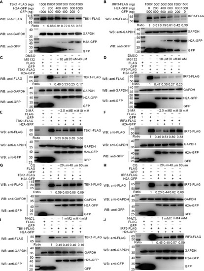
Zebrafish histone H2A degrades TBK1 and IRF3 via the lysosomal pathway. (A) Effect of zebrafish histone H2A on the protein expression of TBK1. (B) Effect of zebrafish histone H2A on the protein expression of IRF3. For (A, B), EPC cells seeded in six-well plates were transfected with various indicated plasmids and DNA concentration. After 48 h post-transfection, cell lysates were analyzed by Western blotting using the indicated Abs. (C) Effect of MG132 on the histone H2A-mediated protein degradation of TBK1. (D) Effect of MG132 on the histone H2A-mediated protein degradation of IRF3. (E) Effect of 3-MA on the histone H2A-mediated protein degradation of TBK1. (F) Effect of 3-MA on the histone H2A-mediated protein degradation of IRF3. (G) Effect of CQ on the histone H2A-mediated protein degradation of TBK1. (H) Effect of CQ on the histone H2A-mediated protein degradation of IRF3. (I) Effect of NH4Cl on the histone H2A-mediated protein degradation of TBK1. (J) Effect of NH4Cl on the histone H2A-mediated protein degradation of IRF3. For (C–J), EPC cells seeded in six-well plates were transfected with various indicated plasmids and DNA concentration. After 48 h post-transfection, cells were treated with DMSO, MG132, 3-MA, CQ or NH4Cl with indicated concentration for 6 h. Following this, cell lysates were analyzed by Western blotting using the indicated Abs. The expression ratio for TBK1 or IRF3 protein was quantified by Quantity One. All experiments were repeated for at least three times with similar results. |