|
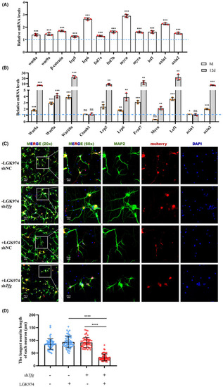
Wnt signaling is activated in response to TFG deficiency. (A) qRT‐PCR‐based validation of the key molecule in Wnt signaling in tfg morphants and control morphants at 3 dpf. (mean ± SD; two‐tailed unpaired t‐test, ***p < 0.001; n = 3). (B) qRT‐PCR‐based validation of the key molecule in Wnt signaling in primary cultured neurons on day 8 and 12 in culture after Tfg knockdown. (mean ± SD; two‐tailed unpaired t‐test; ns, nonsignificant; **p < 0.01; ***p < 0.001; n = 3). (C) Immunofluorescence staining of primary cultured neurons on day 8 after Tfg knockdown and pretreated with LGK974 for 24 h on day 4. Neurites were labeled with MAP2 (green) and the nucleus with DAPI (blue). Scale bars: 20 μm. (D) Quantification of the longest neurite length on day 8 in culture (mean ± SD; two‐tailed unpaired t‐test; ns, nonsignificant; ****p < .0001; n = 60).
|