FIGURE

Figure 3

ID
ZDB-FIG-221104-3
Publication
He et al., 2022 - Anti-Inflammatory and Antioxidant Properties of Physalis alkekengi L. Extracts In Vitro and In Vivo: Potential Application for Skin Care
Other Figures
All Figure Page
Back to All Figure Page
Figure 3

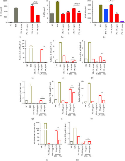
Anti-inflammatory effects of PEs on LPS-stimulated RAW264.7. Cells were pretreated with PEs at the concentrations of 50, 100 μg/mL for 1 h and exposed to 1 µg/mL LPS for another 24 h. After, IL-6 (a), IL-1β (b), and MMP-9 (c) production in the cell culture supernatant were detected by using an enzyme-linked immunosorbent assay (ELISA). The cells were collected and lysed for the measurement of the relative expression of genes involved in immune responses (IL-6 (d), IL-10 (e), IL-α (f), IL-1β (g), TNF-α (h), NOS (i), COX-2 (j), and PGE2 (k)) by qRT-PCR analysis (β-Actin expression was used as an internal control). Each experiment was performed in triplicate. p < 0.05,  ∗∗p < 0.01,  ∗∗∗p < 0.001 compared to the group of LPS-treatment alone.

Expression Data

Expression Detail
Antibody Labeling
Phenotype Data

Phenotype Detail
Acknowledgments
This image is the copyrighted work of the attributed author or publisher, and ZFIN has permission only to display this image to its users. Additional permissions should be obtained from the applicable author or publisher of the image. Full text @ Evid. Based Complement. Alternat. Med.