Fig. 13
- ID
- ZDB-FIG-220801-255
- Publication
- Eugenin von Bernhardi et al., 2022 - A versatile transcription factor: Multiple roles of orthopedia a (otpa) beyond its restricted localization in dopaminergic systems of developing and adult zebrafish (Danio rerio) brains
- Other Figures
- All Figure Page
- Back to All Figure Page
|
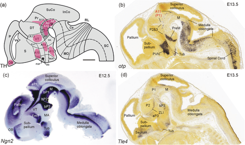
The embryonic mouse basal diencephalon. (a) Schematic sagittal view of the embryonic mouse brain (modified from Vernier & Wullimann, 2009, based on data by Smeets & González 2000; Marín et al., 2005; Björklund & Dunnett, 2007; Vitalis et al., 2000) shows embryonic mammalian dopamine systems. Note position of A11 in the alar plate of prosomere 1 (P1). (b) Embryonic mouse brain sagittal section shows all major otp expression domains (taken from Developmental Mouse Brain Atlas, Allen Institute) except for that in the medial amygdala which is out of section plane. Note strong expression in the alar plate of P1 and sparse expression in the basal plate of P1 through P3. (c) Embryonic mouse brain sagittal section shows expression of bHLH gene ngn2 (modified from Osório et al., 2010). Note the large domain selectively demarcating the region of interest, that is, the basal plate portions of midbrain and P1 through P3 (incl. spear-shaped expression domains flanking in addition the zona limitans intrathalamica between dorsal and ventral thalamus). (d) Embryonic mouse brain sagittal section shows expression domains of the geneTle4 (transducin-like enhancer of split 4; taken from Developmental Mouse Brain Atlas, Allen Institute) which has a potential role in basal diencephalic regions (P1–P3). Scale bar: 500 µm (applies to all panels). Abbreviations: A8-A14, mammalian dopaminergic cell groups (see text). aP1/2, alar plates of prosomeres 1/2; bP1-3, basal plates of prosomeres 1–3; Ce, cerebellum; DT, dorsal thalamus; EmT, eminentia thalami; InCo, inferior colliculus; M, midbrain tegmentum; MA, mammillary hypothalamus; MO, medulla oblongata; OB, olfactory bulb; P, pallium; P1-P3, prosomeres 1–3; POA, anterior preoptic area; Pr, pretectum; PreM, premammillary region; PVN, paraventricular nucleus; RCH, retrochiasmatic area; RL, rhombic lip; S, subpallium; SC, spinal cord; SuCo, superior colliculus; Tub, tuberal hypothalamus; Ve (telencephalic), ventricle; VT, ventral thalamus; ZLI, zona limitans intrathalamica |

