
Fig. 6
- ID
- ZDB-FIG-220706-34
- Publication
- Sarohi et al., 2022 - Comprehensive Mapping and Dynamics of Site-Specific Prolyl-Hydroxylation, Lysyl-Hydroxylation and Lysyl O-Glycosylation of Collagens Deposited in ECM During Zebrafish Heart Regeneration
- Other Figures
- All Figure Page
- Back to All Figure Page
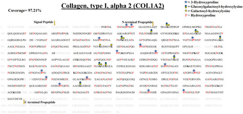
Comprehensive map of COL1A2 of ECM of WT zebrafish heart. It presents proline/lysine hydroxylation sites and lysine glycosylation sites. Representation of PTM sites is similar to COL1A1a and COL1A1b PTM maps. Peptides identified in proteomics analysis are shown in black color and unidentified peptides are shown in grey color. Total 97.21% sequence coverage of COL1A2 is detected (considering the matured form of COL1A1b). The signal peptide is 22 amino acids (1–22) long. N terminal propeptide (23–68) and C terminal propeptide (1,109–1,352) cleavage sites are marked with dark yellow arrows. Red bold “P” with blue star represents 3-HyP and red “P” represents 4-HyP. Hydroxylysine is represented with bold “K” and yellow and blue circle represents the lysine O-glycosylation. A summary of these site-specific PTMs of COL1A2 is presented in Table 1, and all the PSMs for O-glycosylated lysine and 3-hydroxyproline sites are provided in Supplementary Figure S2.53–S2.83. |