
Fig. 1
- ID
- ZDB-FIG-220706-29
- Publication
- Sarohi et al., 2022 - Comprehensive Mapping and Dynamics of Site-Specific Prolyl-Hydroxylation, Lysyl-Hydroxylation and Lysyl O-Glycosylation of Collagens Deposited in ECM During Zebrafish Heart Regeneration
- Other Figures
- All Figure Page
- Back to All Figure Page
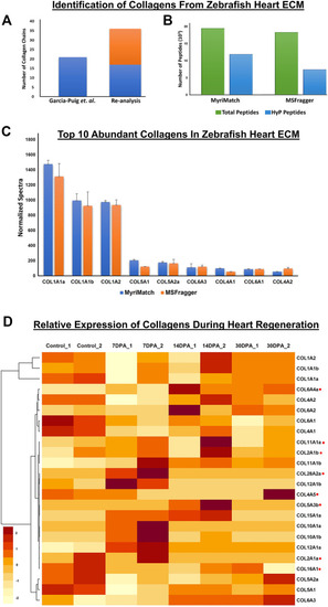
Identification of collagen chains and their relative abundances in zebrafish heart ECM: (A) Total number of collagen chains (Cabral et al., 2007) identified previously by Garcia-Puig et al. compared to (Padmanabhan Iyer et al., 2016) number of collagen chains identified in our analysis. (B) depicts inclusion of hydroxyproline (HyP) modification in the database search by MyriMatch and MSFragger resulting in identification of almost 61.12 and 40.46% (summed number from all the raw *.pepXML files used for database search) new unique peptides. This strategy yielded more no. of peptide identification resulting in a higher number of collagen chain identification from the same dataset. (C) Top 10 abundant collagen chains deposited in the zebrafish heart ECM were identified by two different search engines MyriMatch and MSFragger respectively. (D) Relative abundances of different collagen chains during zebrafish heart regeneration are shown by the heatmap. Light yellow represents the lower value (−2) and dark red represents the higher value (+2) in the row. Normalized spectral count values have been used to generate the heatmap (considering ≥ 3 spectral counts per chain). collagen chains marked with red (.) dots are quantitated during regeneration in re-analysis for the first time (DPA = day post amputation). |