FIGURE

Figure 6

ID
ZDB-FIG-220616-7
Publication
Palominos et al., 2022 - The Olfactory Organ Is a Unique Site for Neutrophils in the Brain
Other Figures
All Figure Page
Back to All Figure Page
Figure 6

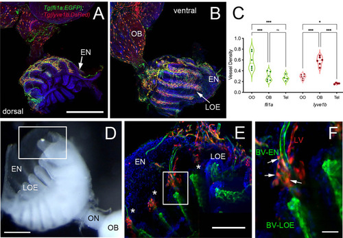
Blood (BV) and Lymphatic (LV) Vasculature wrap the olfactory organs (OO). (A, B) Whole mount Tg(fli1a:EGFP;lyve1b:DsRed) of the adult OO connected to OB with BV (fli1a:EGFP, green) and LV (lyve1b:DsRed, red). Dorsal (A) and ventral (B) views; DAPI (blue), Scale Bars: (A, B) = 200 μm. (C) BV (red) and LV (green) density is greater in olfactory system (OS = OE and OB) than telencephalon (Tel); (SE, P-value <0.05, unpaired t-test; n = 6 adult brains. One-way ANOVA, Tukey multiple comparison test, P < 0.05). Representative images selected from detailed analysis of least 6 brains. (D) Transmitted light image of fixed whole mount OO. Boxed area corresponds to area where LOE connects with EN. Scale bar = 100 μm, (E). The LV (red) meet the BV (green) at the distal tips of each lamellae (boxed area; asterisks). Scale bar = 100 μm, (F). Cells express both lyve1b:DsRed and fli1a:EGFP (arrows). Scale bar = 25 μm. (A, B), (E, F): Analysis of 9 brains. Two-way ANOVA, Tukey’s multiple comparison test, p < 0.05). (*P, 0.05, ***P, 0.001, ns: non significant).

Expression Data

Expression Detail
Antibody Labeling
Phenotype Data

Phenotype Detail
Acknowledgments
This image is the copyrighted work of the attributed author or publisher, and ZFIN has permission only to display this image to its users. Additional permissions should be obtained from the applicable author or publisher of the image. Full text @ Front Immunol