|
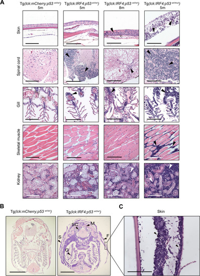
<italic>IRF4</italic>-driven zebrafish tumors recapitulate invasive human T-cell lymphoma.A Histopathological examination of H&E-stained tissue sections of representative samples from control Tg(lck:mCherry;p53wt/wt) (n = 8), Tg(lck:IRF4;p53wt/wt) (n = 8) and Tg(lck:IRF4;p53wt/mut) (n = 4) zebrafish at 5 or 8 months. Similar findings were observed in multiple independent animals as shown for each sample. Tumor cells are indicated by white or black arrowheads. Scale bar = 100 μm. B Transverse sections of representative samples from Tg(lck:mCherry;p53wt/wt) (n = 8) and Tg(lck:IRF4;p53wt/wt) (n = 8). F, fin; G, gill; M, muscle; S, skin; and SC, spinal cord. Similar findings were observed in multiple independent animals as shown for each sample. Scale bar = 2 mm. C High magnification of skin lesions from Tg(lck:IRF4;p53wt/wt) fish. Scale bar = 100 μm.
|