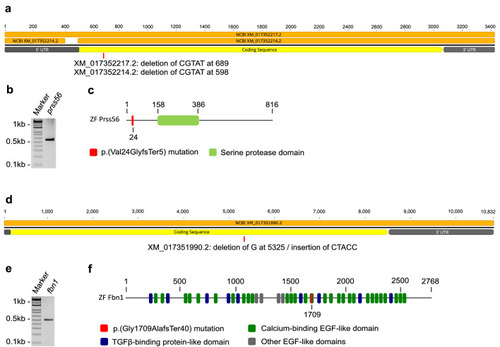
Expression of prss56 and fbn1 and characteristics of the induced mutations. Schematic overview of predicted cDNA and protein structures and induced mutations in mutant zebrafish. (a) Schematic illustration of the cDNA structure of prss56. The two isoforms XM_017352214.2 and XM_017352217.2 are depicted in orange and the induced mutations are shown for each isoform at the approximate location. (b) Agarose gel loaded with reverse transcriptase polymerase chain reaction product (primer set 4) of isolated RNA extracted from 6 mpf zebrafish eyes, confirming ocular expression of prss56. See Supplementary Table S1 and Supplementary Fig. S3 for more information on primers. (c) Schematic illustration of the Prss56 protein structure and induced mutation (in red). (d) Schematic illustration of the cDNA structure of fbn1. (e) Agarose gel loaded with reverse transcriptase polymerase chain reaction product of isolated RNA extracted from 6 mpf zebrafish eyes, confirming ocular expression of fbn1. See Supplementary Table S1 for primers. (f) Schematic illustration of the Fbn1 protein structure and induced mutation (in red).
|

