
Figure 2
- ID
- ZDB-FIG-220131-29
- Publication
- Westphal et al., 2022 - Wnt/β-catenin signaling promotes neurogenesis in the diencephalospinal dopaminergic system of embryonic zebrafish
- Other Figures
- All Figure Page
- Back to All Figure Page
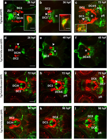
Activity domains of Wnt/β-catenin signaling in relation to TH-immunoreactive cells in the ventral diencephalon and hypothalamus. (a–l) Wnt/β-catenin-reporter Tg(top:dGFP) (a–c) and Tg(7xtcf-Xla.siam:GFP) (d–l) zebrafish embryos were stained by double immunofluorescence for TH-immunoreactive cells (red) and GFP-immunoreactive cells (green) at indicated stages. Dorsal views of the ventral diencephalon/hypothalamus region. Confocal image stacks were recorded and images show 1.2 µm single optical sections of Z-stacks containing TH-immunoreactive cells. DA neuron groups DC2 and DC4 of the ventral diencephalon, DC3 of the medial hypothalamus and DC5 and DC6 of the hypothalamus are labeled. (e,f) Two optical sections of a single 48 hpf embryos at 33.02 µm distance from dorsal (e) to ventral (f). (g–i) Three optical section of single 72 hpf embryo at (g–i) 16.51 µm and (i,j) 11.43 µm distances, with the dorsalmost section shown in (g). (j–l) Three optical section of a single 96 hpf embryo at (j–k) 11.43 µm and (k–l) 13.47 µm distances, with the dorsalmost section shown in (j). (d–l) Z-stacks are included in Supplementary Information as Supplementary Video 4 (g), Supplementary Video 5 (e,f), Supplementary Video 6 (g–i) and Supplementary Video 7 (j–l). Inserts in (a–c) show higher magnifications of boxed areas in (a–c). Scale bar in (a) is 20 µm for (a–c) and in (d,g,j) is 50 µm for (d–l) Scale bars in inserts are 5 µm for (a–c). DC diencephalon, H hypothalamus, pR posterior recess, ZLI Zona limitans intrathalamica. |