|
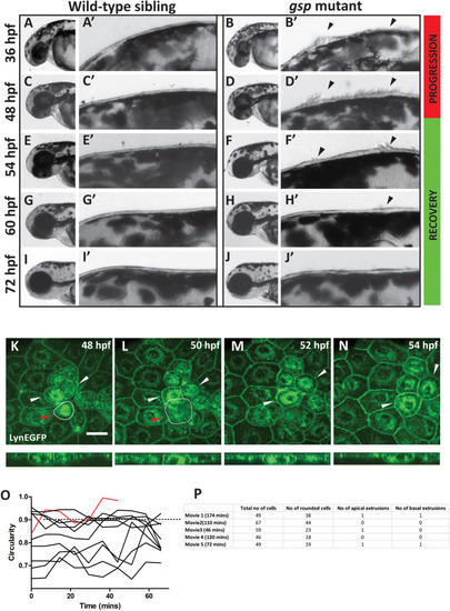
Regression of peridermal cell rounding phenotype occurs through reintegration of rounded cells.DIC images of individual wild-type sibling (A, C, E, G, I) and gsp mutant (B, D, F, H, J) embryos imaged successively at 36, 48, 54, 60, and 72 hpf, respectively. A’–J’ represent the corresponding enlarged images of dorsal head epidermis. Arrowheads indicate regions showing cell rounding. Note that cell rounding increases until 48 hpf (progression) and decreases subsequently (recovery) to finally cease by 72 hpf. Confocal time lapse images of dorsal head epidermis of cldnB:lynEGFP embryos injected with myoVb splice site morpholino (myoVb MO) during the recovery period at 48 (K), 50 (L), 52 (M), and 54 (N) hpf along with respective orthogonal projections. All cell borders as well as several intracellular membrane compartments are marked by lynEGFP (green). White arrows represent reintegrated cells, showing progressive cell shape change from round to polygonal. Red arrow and white dotted outline indicate a rounded cell undergoing apical extrusion. Scale bar = 50 μm. Traces showing changes in cell circularity values (O) for ten randomly selected cells in the above movie. Red trace represents extruding cell. Dotted line represents cell circularity = 0.9, which was used as the benchmark for classifying cells as rounded. A table (P) showing total number of cells, number of rounded cells, apical extrusions and basal extrusions (delamination) in live confocal movies of myoVb morphant embryos made between 50-53hpf. The duration of the movie is indicated in parentheses.
|

