FIGURE

Fig. 3

ID
ZDB-FIG-210512-26
Publication
Zhang et al., 2021 - Three-dimensional microscopy and image fusion reconstruction analysis of the thyroid gland during morphogenesis
Other Figures
All Figure Page
Back to All Figure Page
Fig. 3

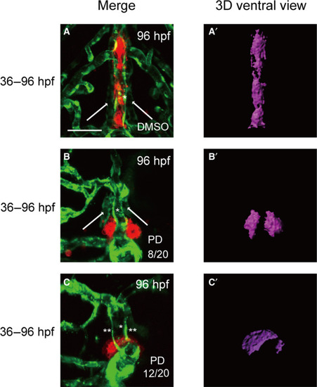
Treatment of embryos with the FGFR1‐selective inhibitor PD166866 from 36 to 96 hpf caused severe defects in thyroid morphology and volume and resulted in abnormal or absent in HAs. All embryos shown are oriented anterior to the top in the ventral views. (A) An analysis of DMSO‐treated embryos showed normal vascular development with normal thyroid morphology at 96 hpf. (B) Treatment with PD 166866 from 36 to 96 hpf caused a reduction in tg signal intensity and resulted in abnormal HAs (stubby and malformed) in 8 of 20 embryos. (C) The absence of HAs was observed in 12 of 20 embryos during 36–96 hpf drug treatment, and this occurred along with abnormal thyroid expansion. The percentage of abnormal or absent HAs in PD 166866‐treated embryos is presented in the lower right corner of B and C. Arrow: HA. *: VA. **: the absence of HA. Scale bar: 50 μm.

Expression Data

Expression Detail
Antibody Labeling
Phenotype Data

Phenotype Detail
Acknowledgments
This image is the copyrighted work of the attributed author or publisher, and ZFIN has permission only to display this image to its users. Additional permissions should be obtained from the applicable author or publisher of the image. Full text @ FEBS Open Bio