
Fig. 2
- ID
- ZDB-FIG-210512-25
- Publication
- Zhang et al., 2021 - Three-dimensional microscopy and image fusion reconstruction analysis of the thyroid gland during morphogenesis
- Other Figures
- All Figure Page
- Back to All Figure Page
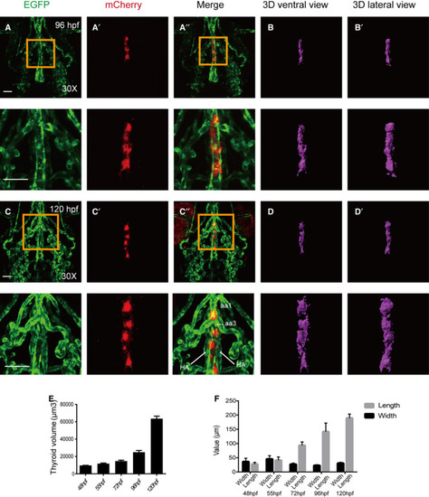
Confocal microscopy images of thyroid expansion from 96 to 120 hpf with 3D simulated images. All embryos shown are oriented anterior to the top in the ventral views. (A‐B) the thyroid is slimmer and shaped more like a long column at 96 hpf. (C‐D) Expansion of thyroid tissue moving away from the heart along the pharyngeal midline occurs at 120 hpf. (E) Thyroid volume expands gradually from 48 to 72 hpf and more drastically from 96 to 120 hpf. (F) Length increases gradually from 48 to 120 hpf, while the width remains almost constant, and this is consistent with the volume changes at the corresponding time point. Magnified views of the yellow box are presented below the corresponding images. * aa1: aortic arch artery 1; *** aa3: aortic arch artery 3. Scale bar: 50 μm. The error bars represent SEM. Data are representative of three independent experiments with similar results. |