|
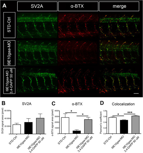
3,4-Diaminopyridine phosphate administration increased pre- and postsynaptic signal co-localization. (A) Representative confocal fluorescence maximum projection images of SV2A (green) and α-BTX (red) signal in 5 spinal hemisegments and somites in embryos at 48 hpf. The images are representative of those found in n = 10 embryos for each experimental group (controls, morphants and 3,4-DAP treated morphants), during 3 distinct experiments. Scale bar = 25 µm. (B–D) Quantification of the SV2A (B) green signal, the α-BTX (C) red signal, and the co-localization signal (D). Error bars are SEMs. (For interpretation of the references to colour in this figure legend, the reader is referred to the web version of this article.)
|