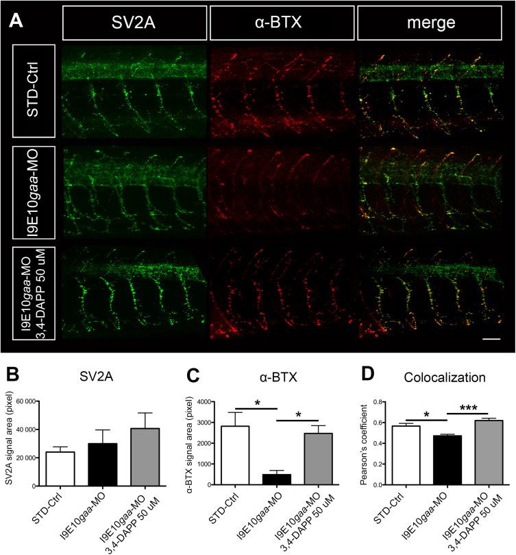
Fig. 2 3,4-Diaminopyridine phosphate administration increased pre- and postsynaptic signal co-localization. (A) Representative confocal fluorescence maximum projection images of SV2A (green) and α-BTX (red) signal in 5 spinal hemisegments and somites in embryos at 48 hpf. The images are representative of those found in n = 10 embryos for each experimental group (controls, morphants and 3,4-DAP treated morphants), during 3 distinct experiments. Scale bar = 25 µm. (B–D) Quantification of the SV2A (B) green signal, the α-BTX (C) red signal, and the co-localization signal (D). Error bars are SEMs. (For interpretation of the references to colour in this figure legend, the reader is referred to the web version of this article.)
Image
Figure Caption
Figure Data
Acknowledgments
This image is the copyrighted work of the attributed author or publisher, and
ZFIN has permission only to display this image to its users.
Additional permissions should be obtained from the applicable author or publisher of the image.
Full text @ Biomed. Pharmacother.

