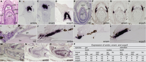
(A) Hematoxylin-eosin (H&E) staining of a developing gar tooth immediately after acrodin formation. Scale bar, 50 μm. (B–D) Our in situ hybridization (ISH) analysis reveals expression of gar ambn (B), enam (C), and scpp5 (D) in IDE cells during the matrix formation stage of acrodin formation (closed arrowheads). (E) H&E staining of a developing gar tooth forming collar enamel. Scale bar, 50 μm. (F–H) ISH analysis reveals expression of gar ambn (F), enam (G), and scpp5 (H) in IDE cells during the secretory stage of collar enamel formation (closed arrowheads). (I) H&E staining of a gar scale forming ganoin. Ganoin was lost during decalcification. Scale bar, 50 μm. (J–L) ISH analysis reveals expression of gar ambn (J), enam (K), and scpp5 (L) in IGE cells during the secretory stage of ganoin formation (closed arrowheads). Brown pigments are shown by asterisks. (M) H&E staining of a developing zebrafish tooth. Scale bar, 50 μm. (N and O) ISH analysis reveals expression of zebrafish ambn (N) in IDE cells (a closed arrowhead) and enam (O) in matrix-formation stage IDE cells and odontoblasts (closed arrowheads). (P) Summary of matrix SCPP gene expression in mineralized tissues. In gar scales, relative expression levels were determined by RNA-seq analysis (Table S1) and the highest expression level of scpp5 among all SCPP genes is shown as “++.” Expression of zebrafish scpp5 (Kawasaki et al., 2017) and stages of hypermineralized tissue formation in gar and teleosts were described previously (Sasagawa, 1995, 1997; Sasagawa and Ishiyama, 2005; Sasagawa et al., 2008, 2013). See Figure S2A for negative controls and more results. Abbreviations: acr, acrodin; bo, bone; cem, collar enamel; de, dentine; ga, ganoin; ide, inner dental epithelial cells; ige, inner ganoin epithelial cells; od, odontoblasts.
|
