FIGURE

Figure 5

ID
ZDB-FIG-210301-56
Publication
Müller-Deile et al., 2021 - Novel diagnostic and therapeutic techniques reveal changed metabolic profiles in recurrent focal segmental glomerulosclerosis
Other Figures
All Figure Page
Back to All Figure Page
Figure 5

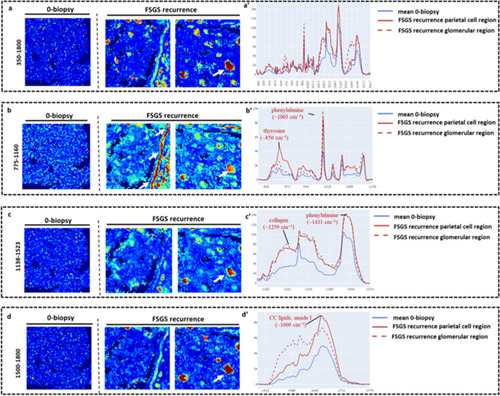
Machine learning reveals anomalies in Raman spectroscopy maps between 0-biopsy and FSGS recurrence. Raman spectra of glomeruli from the 0-biopsy and two glomeruli from the biopsy with FSGS recurrence were visualized, whereby the intensity and color range cover the degree of the anomaly. Areas of focal glomerular lesions as well as parietal epithelial cells in the Bowman capsule are highlighted as an anomaly in the FSGS samples. (a) Visualization of the occurring anomaly using the entire spectrum. (bd) Consideration of only parts of the spectrogram that have been baseline corrected individually. Especially in the range of 775–1160 cm−1 (b) there were significant differences in FSGS glomeruli comparison to spectra from glomeruli of the 0-biopsy in focal areas as well in parietal cell region of the bowman capsule. Likewise, a similar effect was observed for the wavelength range 1138–1500 cm−1 (c) and 1500–1800 cm−1 (d). (a′d′) Raman spectra for the anomalies with confidence > 80% in FSGS recurrence of the parietal cell region (red line) and the glomerular region (spotted red line) compared to 0-biopsy (blue line) for the entire spectrum (a′) and divided in wavelength range of 775–1160 cm−1 (b′) 1138–1523 cm−1 (c′) as well as 1500–1800 cm−1 (d′).

Expression Data

Expression Detail
Antibody Labeling
Phenotype Data

Phenotype Detail
Acknowledgments
This image is the copyrighted work of the attributed author or publisher, and ZFIN has permission only to display this image to its users. Additional permissions should be obtained from the applicable author or publisher of the image. Full text @ Sci. Rep.