FIGURE

Fig. 9

ID
ZDB-FIG-200730-5
Publication
Hall et al., 2020 - In vivo cell biological screening identifies an endocytic capture mechanism for T-tubule formation
Other Figures
All Figure Page
Back to All Figure Page
Fig. 9

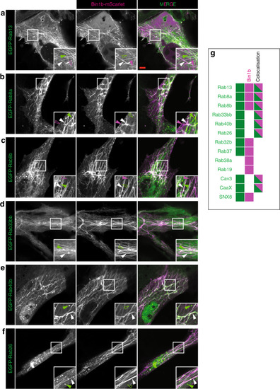
An in vitro model of T-tubule formation.

(a) Rab13, (b) Rab8a, (c) Rab8b, (d) Rab33bb, (e) Rab40b, (f) Rab26, (g) summary graphic showing cells that possess tubules with Bin1b only (magenta), marker only (green) and tubules where bin1b colocalizes with the marker (dual colour; (see also Supplementary Fig. 9). Images are representative of three individual cells imaged within the same experiment.

Expression Data

Expression Detail
Antibody Labeling
Phenotype Data

Phenotype Detail
Acknowledgments
This image is the copyrighted work of the attributed author or publisher, and ZFIN has permission only to display this image to its users. Additional permissions should be obtained from the applicable author or publisher of the image. Full text @ Nat. Commun.