
Fig. 6
- ID
- ZDB-FIG-200306-121
- Publication
- Nimura et al., 2019 - Role of Reelin in cell positioning in the cerebellum and the cerebellum-like structure in zebrafish
- Other Figures
- All Figure Page
- Back to All Figure Page
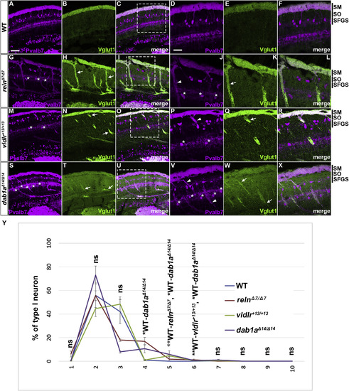
Aberrant positioning of type I neurons in adult zebrafish reln, vldlr, and dab1a mutants. (A–X) Cross sections of the brain from adult (90–150 dpf) WT (A-F, n= 9) and relnΔ7/Δ7 (G-L, n= 5), vldlr+13/+13 (M-R, n= 5), and dab1aΔ14/Δ14 (S-X, n= 4) mutant zebrafish were stained with anti- Pvalb7 (magenta) and anti-Vglut1 (green). Typical tectum images are shown. (D-F, J-L, P-R, V-X) High magnification images of the boxes in C, I, O, and U. Typical images are shown. Pvalb7+ type I neurons were located in a restricted area of the stratum fibrosum et griseum superficiale (SFGS) layer in the tectum in WT (A, D), but were ectopically localized in reln, vldlr, and dab1a mutants (indicated by asterisks in G, J, M, P, S, and V). Dendrites of some type I neurons were misoriented (indicated by arrowheads in J, P, and V). The Vglut1+ axons of granule cells (GCs) in the tectum project to dendrites of type I neurons in the stratum marginale (SM) in WT (B, E). These axons projected to ectopic type I neurons in reln, vldlr, and dab1a mutants (indicated by arrows in H, K, N, Q, T, and W). The stratum opticum (SO) is located between the SM and the SFGS. (Y) Position of type I neurons in WT (n= 9), relnΔ7/Δ7 (n= 4), vldlr+13/+13 (n= 4), and dab1aΔ14/Δ14 (n= 4). The tectum region was divided into ten domains from the pia to ventricle. The positions of the type I neurons were determined by measuring the distance between the pia and the soma of type I neurons. Pvalb7+ cells located in the vicinity of the ventricular zone that had a morphology different from type I neurons were excluded. Average and standard deviation are indicated in the graph. *p < 0.05; **p < 0.01; ***p< 0.001; ns not significant (Dunn’s multiple comparison test). Scale bars: 20 μm in A (applies to A-C, G-I, M-O, and S–U); 40 μm in D (applies to D-F, J-L, P-R, and V-X). |
| Genes: | |
|---|---|
| Antibodies: | |
| Fish: | |
| Anatomical Terms: | |
| Stage: | Adult |
| Fish: | |
|---|---|
| Observed In: | |
| Stage: | Adult |
Reprinted from Developmental Biology, 455(2), Nimura, T., Itoh, T., Hagio, H., Hayashi, T., Di Donato, V., Takeuchi, M., Itoh, T., Inoguchi, F., Sato, Y., Yamamoto, N., Katsuyama, Y., Del Bene, F., Shimizu, T., Hibi, M., Role of Reelin in cell positioning in the cerebellum and the cerebellum-like structure in zebrafish, 393-408, Copyright (2019) with permission from Elsevier. Full text @ Dev. Biol.