
Fig. 2
- ID
- ZDB-FIG-200306-117
- Publication
- Nimura et al., 2019 - Role of Reelin in cell positioning in the cerebellum and the cerebellum-like structure in zebrafish
- Other Figures
- All Figure Page
- Back to All Figure Page
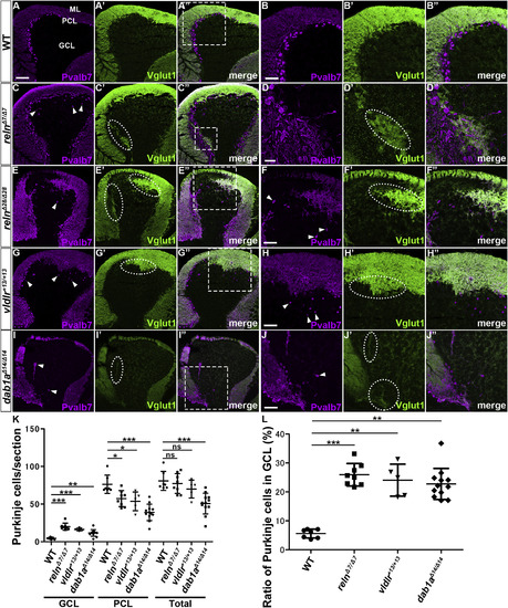
Aberrant positioning of Purkinje cells (PCs) in adult zebrafish reln, vldlr, and dab1a mutants. (A–J) Sagittal sections of the brain from adult (90–150 dpf) wild-type (WT, A-A″, n= 6), and reln (C–C″, E-E″, n= 5 for relnΔ7/Δ7 and n= 5 for relnΔ28/Δ28), vldlr+13/+13 (G-G″, n= 5), and dab1aΔ14/Δ14(I–I″, n= 8) mutant zebrafish were stained with anti-parvalbumin7 (Pvalb7, magenta) and anti-Vglut1 (green) antibodies. Typical cerebellum images are shown. (B–B″, D-D″, F–F″, H–H″, J-J″) High magnification images of the boxes in A", C", E", G", and I". Ectopic Purkinje cells (PCs) are indicated by arrowheads. Axonal projections of granule cells (GCs) to ectopic PCs are marked by dotted circles. (K) Ectopic PCs. Sagittal sections 14-μm thick were prepared from adult zebrafish WT (n= 7), and relnΔ7/Δ7 (n= 8), vldlr+13/+13 (n= 5), and dab1aΔ14/Δ14 (n= 12) mutant brains. Pvalb7+ PCs in the granule cell layer (GCL) or the Purkinje cell layer (PCL) were counted in every fourth section (14 total sections near the midline of each fish). Average numbers and standard deviations of PCs in the GCL, PCL, or all layers (Total) are shown in the graph. (L) Proportion of PCs in the GCL in WT, and relnΔ7/Δ7, vldlr+13/+13, and dab1aΔ14/Δ14 mutant cerebellum. A greater proportion of the total PCs were located in the GCL in the reln, vldlr, and dab1a mutants than in WT. *p < 0.05; **p < 0.01; ***p< 0.001; ns not significant (Dunnett’s test for K and Dunn’s multiple comparison test for L). Scale bars: 100 μm in A (applies to A-A″, C–C″, E-E″, G-G″, I–I″); 50 μm in B, D, F, H, and J (applies to B–B″, D-D″, F–F″, H–H″ and J-J″, respectively). |
| Genes: | |
|---|---|
| Antibodies: | |
| Fish: | |
| Anatomical Terms: | |
| Stage: | Adult |
| Fish: | |
|---|---|
| Observed In: | |
| Stage: | Adult |
Reprinted from Developmental Biology, 455(2), Nimura, T., Itoh, T., Hagio, H., Hayashi, T., Di Donato, V., Takeuchi, M., Itoh, T., Inoguchi, F., Sato, Y., Yamamoto, N., Katsuyama, Y., Del Bene, F., Shimizu, T., Hibi, M., Role of Reelin in cell positioning in the cerebellum and the cerebellum-like structure in zebrafish, 393-408, Copyright (2019) with permission from Elsevier. Full text @ Dev. Biol.