FIGURE

Figure 3

ID
ZDB-FIG-191230-648
Publication
Koch et al., 2019 - Aspergillus fumigatus establishes infection in zebrafish by germination of phagocytized conidia, while Aspergillus niger relies on extracellular germination
Other Figures
All Figure Page
Back to All Figure Page
Figure 3

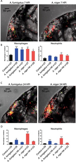
Quantification of leukocyte migration to the hindbrain at early and later stages of infection. (A,C) Representative confocal stacks of Tg (mpeg1:mCherry; mpx:GFP) zebrafish embryos with macrophages and neutrophils labelled in red and green respectively, injected with 150 conidia of A. fumigatus and A. niger at 7 HPI (A) and 24 HPI. (C) Stacks formed the basis of quantification of leukocyte migration to the hindbrain. As Macrophages and conidia were labelled with the same fluorophore, intensity of signal and morphology was used to distinguish conidia from macrophages. (B,D) Quantification of leukocyte migration in the hindbrain of embryos injected with A. fumigatus or A. niger, live or heat-killed (HK) conidia, compared to control injections (2% PVP/PBS) at 7 HPI (B) and 24 HPI. (D) *p ≤ 0,05; **p ≤ 0,01; ***p ≤ 0,001; ****p ≤ 0,0001 by one-way ANOVA with Dunnett’s multiple comparisons test, n = 21–30 per group from 3 biological replications. The quantifications represent a part of a larger data-set which is presented in full in Supplemental Fig. 1. The statistical analysis presented is based on the full dataset.

Expression Data

Expression Detail
Antibody Labeling
Phenotype Data

Phenotype Detail
Acknowledgments
This image is the copyrighted work of the attributed author or publisher, and ZFIN has permission only to display this image to its users. Additional permissions should be obtained from the applicable author or publisher of the image. Full text @ Sci. Rep.