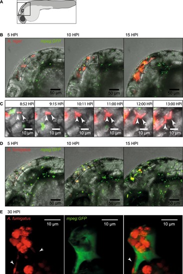
Confocal microscopy captures different infectious development of A. niger and A. fumigatus. (A) Schematic drawing of the embryo and the area imaged in the figure. (B) Still images from confocal time-lapse microscopy (Supplementary Movie SM1), depicting the typically rapid development of A. niger infection. Images from 5, 10 and 15 HPI of 150 conidia of A. niger in Tg(mpeg1:EGFP). At 15 HPI several conidia has germinated and progressive hyphal growth is evident. (C) Digitally magnified still images from Supplementary Movie SM1, aiming to capture the events of extracellular germination and hyphal growth of two separate conidia from approximately 9 HPI to 13 HPI. (D) Still images from confocal time-lapse microscopy (Supplementary Movie SM3), depicting the development of A. fumigatus infection at 5, 10 and 15 HPI, after injection of 150 conidia of A. fumigatus in Tg(mpeg1:EGFP). No germination events were detectable at the end of the time-lapse (Supplementary Movie SM3). (E) High magnification confocal microscopy showing a cluster of A. fumigatus conidia in the hindbrain of a Tg(mpeg1:EGFP) zebrafish embryo acquired at 30 hours post infection of 150 conidia. In the left panel, showing only the red fluorescence channel, several hyphae could be seen protruding from the cluster of several conidia (arrowheads). In the central panel, showing the green channel the macrophage can be seen. In the right panel, overlaying the green fluorescence channel it was clear that the cluster of conidia had been phagocytized and were contained within a macrophage. One growing hyphae could be seen to stretch the membrane of the macrophage (arrowhead). (B) White box insert in 10 HPI image represents the magnified area in image panel (C).
|
