FIGURE

Figure 6

ID
ZDB-FIG-191230-1502
Publication
Collodet et al., 2019 - AMPK promotes induction of the tumor suppressor FLCN through activation of TFEB independently of mTOR
Other Figures
All Figure Page
Back to All Figure Page
Figure 6

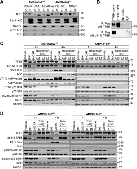
Pharmacological activation of AMPK leads to dephosphorylation and nuclear localization of TFEB. A) AMPKα1α2+/+ or AMPKα1α2−/− MEFs were treated with vehicle (DMSO), 10 µM 991, or 2 mM AICAR for 1 h. Cytoplasmic and nuclear fractions were prepared and lysates (20 µg) from the fractions were used for Western blot analysis of the indicated proteins. B) COS1 cells were transfected for 48 h with no vector (negative control) or the vector containing Flag-tagged TFEB WT or S142A mutant construct. The WT or mutant TFEB was immunoprecipitated (IP) from cell lysate (500 μg) with 1 μg of anti-Flag antibody and the immunoprecipitates were subjected to Western blot analysis, with total or phospho-S142 (pS142) TFEB antibodies. Images are representative of n = 2. C) MEFs were treated with AMPK activators (10 µM 991 or 2 mM AICAR) or various concentrations of mTOR inhibitor rapamycin (0.05, 0.1 and 0.5 µM) for 1 or 4 h. Cell lysates (20 µg) were subjected to Western blot analysis using the indicated antibodies. D) The experiment was performed as described in C, except Torin-2 (5 or 10 nM) was used as an alternative mTOR inhibitor. Images are representative of n = 2.

Expression Data

Expression Detail
Antibody Labeling
Phenotype Data

Phenotype Detail
Acknowledgments
This image is the copyrighted work of the attributed author or publisher, and ZFIN has permission only to display this image to its users. Additional permissions should be obtained from the applicable author or publisher of the image. Full text @ FASEB J.