
FIGURE
Fig. S1
- ID
- ZDB-FIG-190702-27
- Publication
- Umali et al., 2019 - Loss of foxc1 in zebrafish reduces optic nerve size and cell number in the ganglion cell layer
- Other Figures
- All Figure Page
- Back to All Figure Page
Fig. S1
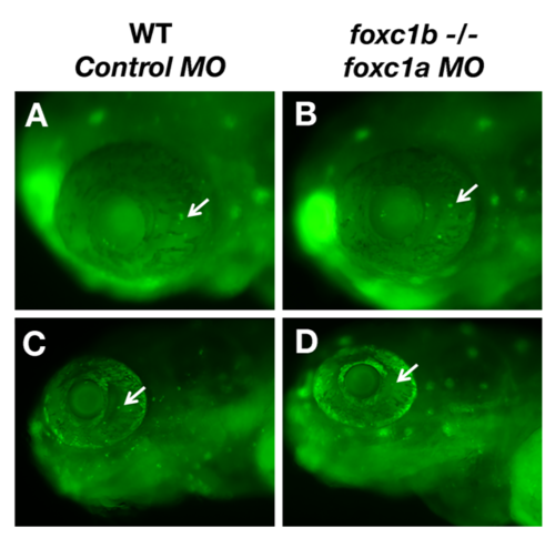
Injection of foxc1a morpholinos into foxc1b -/- mutants does not cause cell death in the eye. A small number of apoptotic cells identified by acridine orange staining are identified in wildtype eyes at 48 hpf and 72 hpf (A and C). No difference in the number of apoptotic cells are observed due to loss of foxc1 function (B and D). |
Expression Data
Expression Detail
Antibody Labeling
Phenotype Data
| Fish: | |
|---|---|
| Knockdown Reagent: | |
| Observed In: | |
| Stage Range: | Long-pec to Protruding-mouth |
Phenotype Detail
Acknowledgments
This image is the copyrighted work of the attributed author or publisher, and
ZFIN has permission only to display this image to its users.
Additional permissions should be obtained from the applicable author or publisher of the image.
Reprinted from Vision Research, 156, Umali, J., Hawkey-Noble, A., French, C.R., Loss of foxc1 in zebrafish reduces optic nerve size and cell number in the ganglion cell layer, 66-72, Copyright (2019) with permission from Elsevier. Full text @ Vision Res.