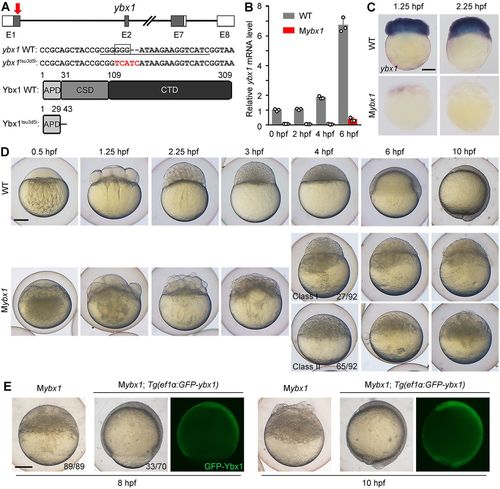
Maternal ybx1 mutant embryos exhibit severe morphological defects. (A) Generation of the ybx1 mutant allele using CRISPR/Cas9. Top: the gRNA target site (red arrow) within the first exon (E1). Grey boxes, white boxes and connecting lines represent the open reading frame, untranslated regions and introns, respectively. Middle: sequences of ybx1 WT and ybx1tsu3d5i alleles near the gRNA target site (underlined) showing the deleted 3 bp (boxed) and the 5-bp insertion (red). Bottom: domains of Ybx1 WT protein and predicted mutant protein. APD, alanine/proline-rich domain; CSD, cold shock domain; CTD, C-terminal domain. (B,C) Loss of ybx1 mRNA in Mybx1 embryos revealed by qRT-PCR (B) and WISH (C). (D) Bright-field images showing the embryonic malformation of Mybx1 mutants in contrast to time-matched WT embryos. (E) Bright-field and fluorescent images of Mybx1 embryos expressing GFP-Ybx1. Mybx1; Tg(ef1α:GFP-ybx1) embryos were obtained by crossing Zybx1; Tg(ef1α:GFP-ybx1) females to WT males. Scale bars: 200 μm.
|

