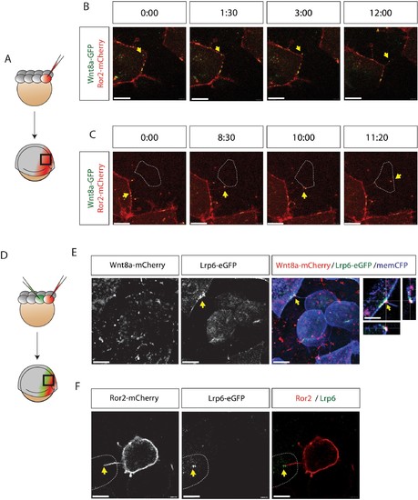
Fig. 5
|
Visualization of Ror2-Wnt8a cytonemal transport and Lrp6 receptor clustering in the living zebrafish. (A) Illustration of the injection procedure to generate single cell clones. (B–C) Time series of a single confocal plane of Ror2-mCherry/Wnt8a-GFP-expressing cells to (B) observe Wnt8a recruitment to the membrane and cytoneme initiation and (C) cytonemal target finding and Ror2/Wnt8a cluster endocytosis into the receiving cell. Optimal microscopical settings were used to allow an improved axial resolution of ca. 250 nm. Single channels in Figure 5—figure supplement 1A,B and Figure 5—video 1. Yellow arrows indicate the Wnt8a/Ror2 cluster and highlights pruning of the cytoneme tip after successful cytonemal delivery. Yellow arrows mark the Wnt8a/Ror2 cluster. (D–F) Wnt8a-mCherry cytoneme leads to Lrp6-GFP accumulation and co-localization at the receiving cell. (D) Illustration of the injection procedure used to generate clones to visualize paracrine signal transport. (E) 3D-stack of a Wnt8a-mCherry cytoneme leading to Lrp6-GFP accumulation and co-localization at the receiving cell. The close-up shows a single plane including orthogonal YZ and XZ views of the cytoneme contact point. (F) Single-plane image of a Ror2-mCherry/Wnt8a cell leading to Lrp6-GFP clustering on the cytoneme contact site. Scale bars = 10 µm; in E = 20 µm. |

