
Fig. 4-S1
- ID
- ZDB-FIG-180724-10
- Publication
- Janjuha et al., 2018 - Age-related islet inflammation marks the proliferative decline of pancreatic beta-cells in zebrafish
- Other Figures
- All Figure Page
- Back to All Figure Page
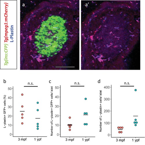
Immune cells infiltrate the islet during development. (a,a’) Confocal image of a juvenile islet. Tg(ins:CFP-NTR) line marks the beta-cells with CFP (green), leukocytes were labelled with the L-plastin antibody (blue) and Tg(mpeg1:mCherry) marks the macrophages (red). (b) Quantification of the percentage of TgBAC(tnfa:GFP) and L-plastin double-positive cells over the total number of L-plastin-positive cells in the islets of TgBAC(tnfa:GFP) animals at 3 mpf (n = 5) and 1 ypf (n = 5). Horizontal bars represent mean values (two-tailed t-test, p>0.05). (c) Quantification of the total number of TgBAC(tnfα:GFP)-positive and L-plastin double-positive cells at 3 mpf (n = 5) and 1 ypf (n = 5). Horizontal bars represent mean values (two-tailed t-test, p>0.05). (d) Quantification of the total number of L-plastin positivecells at 3 mpf (n = 5) and 1 ypf (n = 5). Horizontal bars represent mean values (two-tailed t-test, p>0.05). |