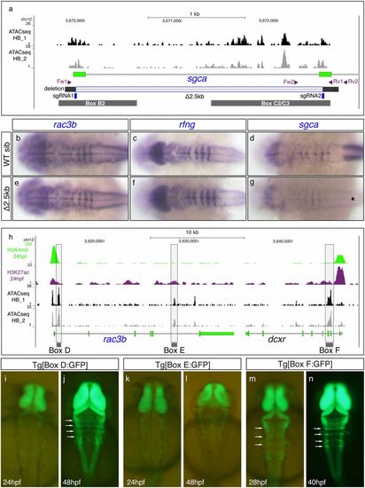
CRISPR-Cas9 deletion of hindbrain boundary cis-regulatory elements revealed the existence of redundant enhancers. (A) Scheme depicting the 2.5kb deletion induced by CRISPR-Cas9 technology (Δ2.5kb; gray-shadowed stretch) containing Box B2 and Box C2/3, along with ATAC-seq profiles from 24 hpf dissected hindbrains (black and gray profiles correspond to two different replicates). The position of the sgRNAs (in blue) used to generate the 2.5kb deletion and the two pairs of primers (in magenta) used for genotyping the mutant line are displayed. (B–G) In situ hybridization analyses of rac3b, rfng, and sgca in WT siblings (B–D) and CRISPR-Δ2.5kb homozygous mutant embryos (E–G) at 22 hpf. Note that rac3b and rfng expression does not considerably change within the hindbrain boundaries between WT and homozygous mutant embryos. sgca expression in the somites is abolished in mutant embryos (black asterisk in G), due to the deletion of the main sgca promoter. All images are dorsal views of flat-mounted hindbrains with anterior to the left. (H) Epigenetic profiles of putative promoters (H3K4me3; green peaks) and active enhancers (H3K27ac; magenta peaks) are shown within the chromosomal region containing rac3b and dcxr along with ATAC-seq signatures (black and gray profiles) from dissected hindbrains at 24 hpf. Three regions associated with the most prominent ATAC-seq peaks were selected (shadowed in gray, Boxes D–F) and each of the fragments was cloned in an enhancer reporter vector to generate a stable transgenic line. (I–N) Dorsal views of embryonic hindbrains from Box D (I and J), Box E (K and L), and Box F (M and N) stable transgenic lines at indicated stages. Note that Tg[Box D:GFP] embryos display GFP expression in the hindbrain boundaries starting at 48 hpf and that Box F is able to drive GFP expression to the hindbrain boundaries before 28 hpf (white arrows in J, M, and N). Box E did not drive GFP to the boundaries. In all images, anterior is at the top. Overall, the expression of the rac3b/rfng/sgca microsyntenic group at the hindbrain boundaries is regulated by multiple enhancers. We have identified at least two early-activated (Boxes C and F) and two late-activated regulatory elements (Boxes B and D).
|

