
FIGURE
Fig. 1
- ID
- ZDB-FIG-180420-5
- Publication
- Vierstraete et al., 2017 - Accurate quantification of homologous recombination in zebrafish: brca2 deficiency as a paradigm
- Other Figures
- All Figure Page
- Back to All Figure Page
Fig. 1
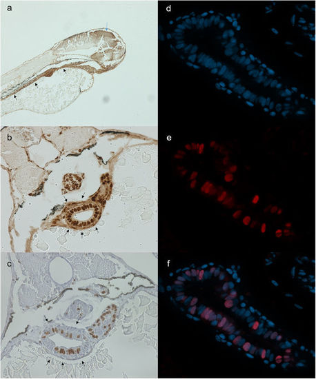
Stainings for PCNA (a,b), BrdU (c) and geminin (d-f). All stainings were performed on 72 hpf embryos. (a) Sagittal section stained for PCNA. Strong staining is found in the tectum (blue arrow) and gastro-intestinal tract (black arrows). (b) Transversal section stained for PCNA. PCNA stains positive in the gut tube, swim bladder and liver. (c) Transversal section stained for BrdU. BrdU intake is prominent in intestinal cells. (d–f) Separate images of nuclear staining (DAPI) (d), mCherry (zGem signal) (e) and overlay (f). (f) mCherry signal is present in a large proportion of intestinal cells. |
Expression Data
| Antibody: | |
|---|---|
| Fish: | |
| Anatomical Term: | |
| Stage: | Protruding-mouth |
Expression Detail
Antibody Labeling
Phenotype Data
Phenotype Detail
Acknowledgments
This image is the copyrighted work of the attributed author or publisher, and
ZFIN has permission only to display this image to its users.
Additional permissions should be obtained from the applicable author or publisher of the image.
Full text @ Sci. Rep.