Fig. 7
- ID
- ZDB-FIG-180126-73
- Publication
- Pei et al., 2016 - Extracellular HSP60 triggers tissue regeneration and wound healing by regulating inflammation and cell proliferation
- Other Figures
- All Figure Page
- Back to All Figure Page
|
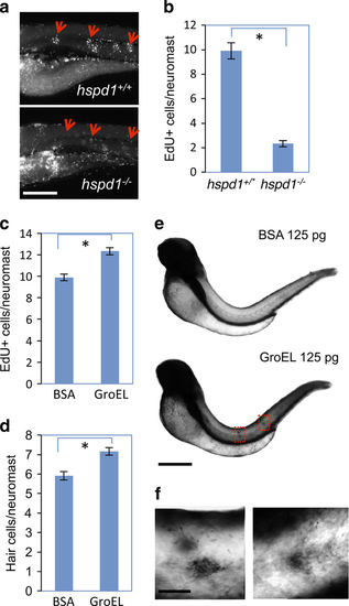
hspd1 is necessary for cell proliferation, and extracellular HSP60 stimulates intracellular hspd1 expression. (a) Impaired proliferation of supporting cell in hspd1 mutants, assayed by EdU labelling analysis. Five-day-old control and hspd1−/− mutant embryos were used for hair cell ablation, EdU labelling and then quantified for cell proliferation. Pictures shown are examples from 24 h post hair cell ablation. Arrows point to the proliferating supporting cells in the lateral line neuromasts. (b) Quantification of the EdU signal. Quantification performed before genotype was determined (n=12, P<0.001). (c) Exogenous GroEL promotes cell proliferation during regeneration. WT embryos at 5 dpf were injected with GroEL, hair cells were ablated and dividing cells labelled with EdU. Data shown were quantified at 24 h post ablation (n=14, P<0.001). (d) Exogenous GroEL promotes supernumerary hair cells. WT embryos at 2 dpf were injected with GroEL into the trunk. Hair cells were counted at 5 dpf by Yopro-1 staining (n=16, P=0.006). (e) GroEL injected in the trunk induces hspd1 expression in the trunk and lateral line neuromasts as revealed by whole-mount in situ hybridisation. WT embryos at 2 dpf were used for 125 pg of GroEL or BSA injection and for analysing hspd1 expression at different time points after the injection by whole-mount in situ hybridiation. Pictures shown are representative examples from 7 h post injection. GroEL-injected embryos showed darker staining in the trunk and lateral line neuromasts. Two red boxes frame the enriched expression of hspd1 in two neuromasts. (f) The magnified images of the two boxed areas of GroEL-injected embryos, revealing the induced hspd1 expression specifically in the neuromasts. Asterisks in b–d indicate a significant difference between the two groups. Bars = 200 μm in a, 500 μm in e and 50 μm in f. dpf, days post-fertilisation. BSA, bovine serum albumin; WT, wild type. |
| Fish: | |
|---|---|
| Condition: | |
| Observed In: | |
| Stage: | Day 6 |

