
Fig. 3
- ID
- ZDB-FIG-180119-29
- Publication
- Eneman et al., 2017 - Pituitary adenylate cyclase-activating polypeptide (PACAP) in zebrafish models of nephrotic syndrome
- Other Figures
- All Figure Page
- Back to All Figure Page
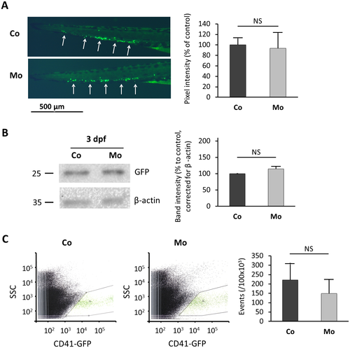
Quantification of thrombocytes in nephrin depleted Tg(cd41:EGFP) transgenic zebrafish. (A) LEFT: GFP-labeled thrombocytes and thrombocyte precursors are formed in the zebrafish caudal hematopoietic tissue (CHT) region (white arrows) at 3 dpf. Representative pictures of the CHT region of a control and a nephrin depleted (100 μM nephrin morpholino) embryo at 3 dpf are shown. No obvious differences in thrombocyte numbers were observed. RIGHT: Pixel intensity was measured using ImageJ software. The graph represents means ± SD from measurements in three embryos per condition. (B) LEFT: Western blot for GFP and β-actin (loading control) was performed using total zebrafish lysates at 3 dpf. A representative blot is shown. RIGHT: Signal intensity was measured using ImageJ software. The graph represents means ± SD from measurements in two repeated experiments. (C) LEFT: Fluorescence-activated cell sorter (FACS) analysis of control zebrafish lysates for CD41 positive cells was performed at 3 dpf. MIDDLE: FACS analysis of morphant zebrafish lysates for CD41 positive cells was performed at 3 dpf. RIGHT: A diagrammatic representation of the number of GFP-positive cells per 100,000 counted cells. For each zebrafish lysate, 500,000 cells were counted and analyzed. Graphs represent means ± SD from three repeated experiments performed in duplicate. Mo, nephrin morpholino injected; Co, control morpholino injected; SCC, side scatter; GFP, green fluorescent protein. |
| Gene: | |
|---|---|
| Fish: | |
| Knockdown Reagent: | |
| Anatomical Terms: | |
| Stage: | Protruding-mouth |
| Fish: | |
|---|---|
| Knockdown Reagent: | |
| Observed In: | |
| Stage: | Protruding-mouth |